37 Thyroid Mass
37.1 Case 1
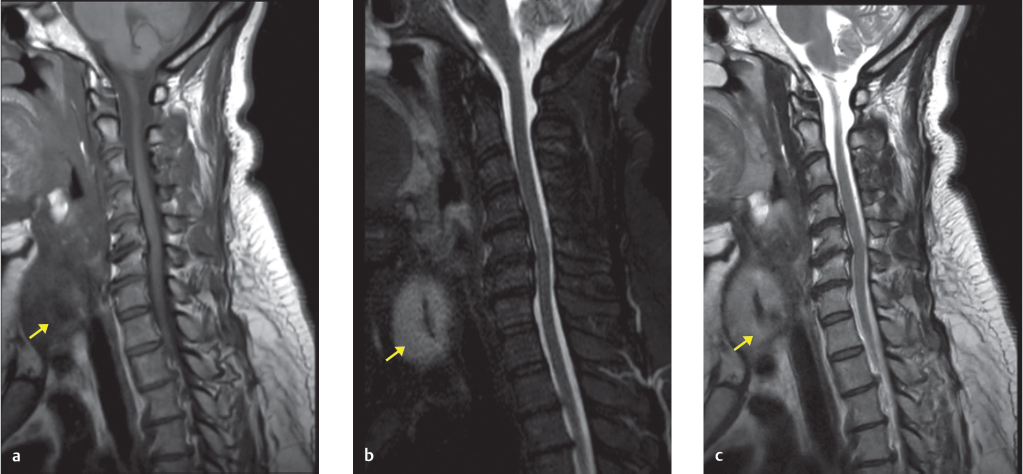
A 49-year-old woman presents with left arm weakness. MRI of the cervical spine without contrast was performed to evaluate for possible cervical nerve root compression (▶ Fig. 37.1).

37.1.1 Imaging Impression
Incidentally detected, 5-cm solid mass in the right lobe of the thyroid gland, which is incompletely imaged on this cervical spine MRI (▶ Fig. 37.2).
37.1.2 Additional Testing Needed: Thyroid Ultrasound
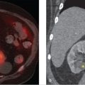
Given that thyroid nodules can have variable signal characteristics depending on their composition and that most MRIs of the cervical spine are not optimized for an accurate diagnosis, thyroid nodules meeting the criteria for size and age based on the ACR Appropriateness Criteria (> 1 cm for individuals younger than 35 years and > 1.5 cm for individuals older than 35 years without history of thyroid cancer or symptomatic thyroid disease) 1 are often further evaluated by thyroid ultrasound. Please see ▶ Table 37.3 for more details. Thyroid ultrasound can provide a higher resolution method to better evaluate the nodules to determine the need for fine needle aspiration (FNA). The thyroid ultrasound is shown in ▶ Fig. 37.3.

37.1.3 Imaging Interpretation
Incidentally detected 4.6-cm thyroid mass is solid, hypoechoic without internal echogenic foci, wider than tall, and fairly well marginated with a slightly lobulated contour.
Based on TI-RADS v2017 criteria, 2 the thyroid nodule scores a total of 6 points and is considered moderately suspicious, meeting the criteria for FNA.
The TI-RADS (Thyroid Imaging Reporting and Data System) v2017 2 grades nodules based on five criteria: nodule composition, nodule border, nodule shape, presence of echogenic foci, and nodule echogenicity; it also assigns points and stratifies nodules to benign (0 points), not suspicious (1–2 points), mildly suspicious (3 points), moderately suspicious (4–6 points), and highly suspicious (≥ 7 points). Please see ▶ Table 37.1 for more details.
Management: ultrasound-guided FNA.
FNA results: adenomatous nodule without malignant features.
Related posts:

Stay updated, free articles. Join our Telegram channel

Full access? Get Clinical Tree


