38

Case 38


Indication: Screening.


History: Unremarkable.


Risk profile: No increased risk.


Age: 57 years.


image


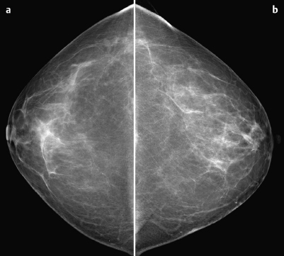
Fig. 38.1 a,b Digital mammography, CC view.


image


Clinical Findings


No findings.


Ultrasound


Unremarkable (not shown).


image


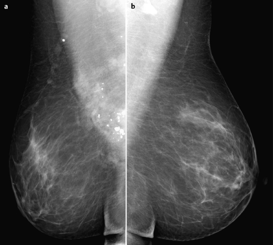
Fig. 38.2a,b Digital mammography, MLO view.


image


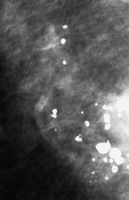
Fig. 38.3 Magnification view of the right breast.


image


image


image


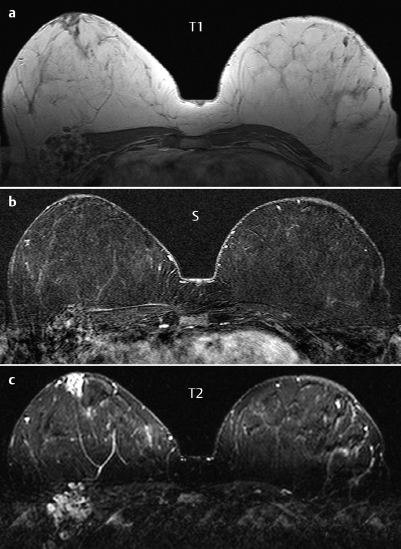
Fig. 38.4a-c Contrast-enhanced MR mammography


image


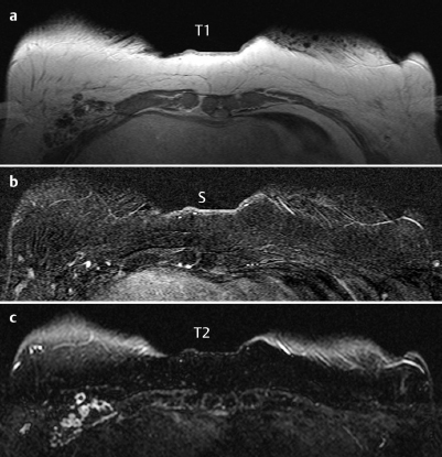
Fig. 38.5a–c Contrast-enhanced MR mammography.


image


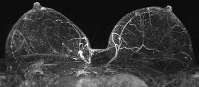
Fig. 38.6 Contrast-enhanced MR mammography. Maximum intensity projection.


image


Please characterize mammography and MRI findings.


What is your preliminary diagnosis?


What are your next steps?


This case illustrates the imaging studies of an asymptomatic woman presenting for stereotactic vacuum core biopsy of “suspicious macro- and microcalcifications” in the lower outer quadrant of the right breast.


Ultrasound


Particularly in the lower outer quadrant of the right breast, there were no findings consistent with the macro- and microcalcifications. US BI-RADS 1.


Mammography


Mammography showed bilaterally symmetric fibroglandular parenchyma, ACR type 2. Multiple monomorphous macrocalcifications were visible in regional distribution in the lower outer quadrant, close to the chest wall. There were no microcalcifications. Mammograms showed no mass or density and no architectural distortion. BI-RADS right 3/left 1. PGMI: CC view G (nipple not in profile); MLO view G (inframammary fold not correctly positioned; superimposition of abdominal wall on parenchyma).


MR Mammography


In precontrast Tl-weighted imaging, MRI documented an area near the chest wall of approx. 3 cm x 3 cm containing multiple foci up to 5 mm in diameter with hypointense signal. After administration of contrast there were individual enhancing spots within this area. Signal in T2-weighted imaging was increased. No other unusual findings.


MRI Artifact Category: 1


MRI Density Type: 1


No evaluable enhancement


MRI score (points)


0


MRI BI-RADS


1


image Preliminary Diagnosis


Hematoma with calcifications.


Differential Diagnosis


Hamartoma, fat necrosis (previous trauma?), hemangioma, angiosarcoma.


BI-RADS Categorization


Clinical Findings


right 1


left 1


Ultrasound


right 1


left 1


Mammography


right 3


left 1


MR Mammography


right 1


left 1


BI-RADS Total


right 3


left 1


Limit of the Requested Stereotactic Vacuum Core Biopsy


In this specific case the stereotactic table (manufacturer: Fischer Imaging) required the use of a diagonal needle angle, in the direction of the chest wall, to access findings close to the chest wall. Therefore, the requested stereotactic biopsy was not performed because of the risk of thoracic injury. At the patient’s request, and despite the BI-RADS 3 categorization of the mammography findings (because of the rare differential diagnosis of angiosarcoma), an open biopsy of this lesion was performed after hook-wire localization.


Macroscopic Findings


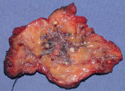
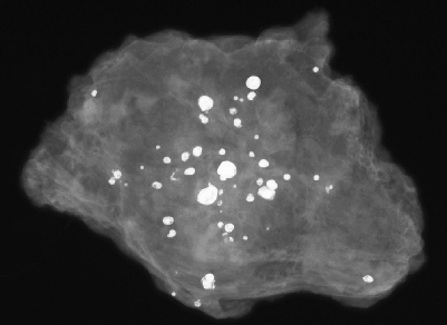
Circumscribed vascular tumor with focal ectasia and partial thrombosis as well as bullet like calcifications. (Fig. 38.7).


image


Fig. 38.7 Excised specimen.


image


Fig. 38.8 Specimen radiography.


Sep 3, 2016 | Posted by in GENERAL RADIOLOGY | Comments Off on 38

Full access? Get Clinical Tree
Get Clinical Tree app for offline access