CASE 38
Clinical Presentation
A 61-year-old man with known coronary artery disease (CAD), prior myocardial infarction, and revascularization is referred for myocardial perfusion imaging to evaluate risk before thoracic surgery. The patient denies any chest pain or shortness of breath. His risk factors include diabetes and hypertension. His medications include metoprolol, enalapril, atorvastatin, aspirin, clopidogrel, and insulin.
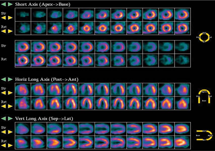
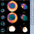
Fig. 38.1
Technique
• The patient had nothing to eat within 4 hours of the test. The β-blocker (metoprolol) was withheld the day of the test. Caffeine-containing beverages were withheld for 24 hours before the test.
• Rest images were acquired 40 minutes after the intravenous injection of 11 mCi of 99mTc-sestamibi. Images were acquired in the supine position with a two-headed gamma camera with step-and-shoot rotation, 32 projections over a 90-degree arc for each head (64 projections over a 180-degree arc), 30 seconds per projection, and a 64 × 64 matrix.
• Vasodilator stress was done with adenosine infused over 4 minutes.
Fig. 38.2
• Heart rate, blood pressure, and 12-lead ECG were recorded at baseline and every minute thereafter during stress.
• A 32 mCi dose of 99mTc-sestamibi was injected during peak stress (at minute 2).
• At 45 minutes after tracer injection, images were obtained with 32 projections over a 90-degree arc for each head of the gamma camera (64 projections over a 180-degree arc), 30 seconds per projection, and a 64 × 64 matrix. Gated images were acquired at 8 frames per cardiac cycle.
Image Interpretation