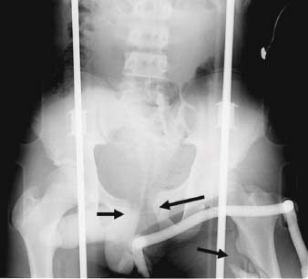
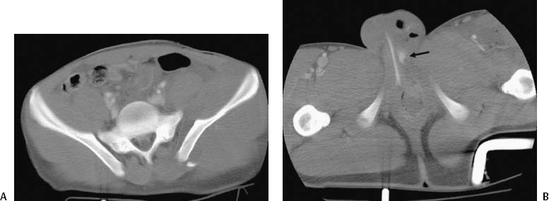
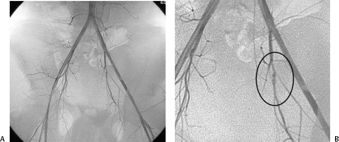
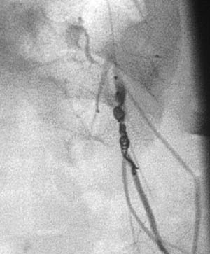
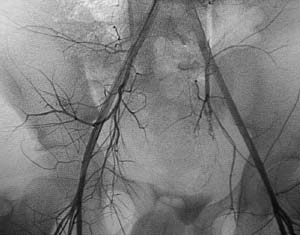
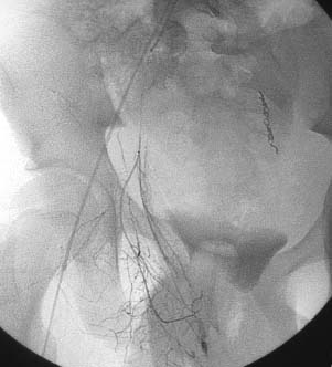
CASE 38 A 14-year-old male presented to the emergency room of an outside hospital after being ejected from an automobile after an accident. He was stabilized with blood products and fluids and underwent plain film and subsequent CT examinations. Figure 38-1 Plain radiograph of pelvic trauma. Plain film of the pelvis with the patient on a backboard shows diastasis of the symphysis pubis and a comminuted left proximal femoral fracture (arrows). Frontal view of the pelvis (Fig. 38-1) obtained on a backboard showed diastasis of the symphysis pubis and a comminuted fracture of the proximal left femur. Views through the pelvis (Fig. 38-2A) showed diastasis of the left sacroiliac joint, an expanding pelvic hematoma, and active bleeding at the base of the penis (Fig. 38-2B). The patient was transferred for urgent pelvic angiography. Pelvic hematoma from arterial hemorrhage after blunt trauma. The right common femoral artery was punctured using the Seldinger technique, and a 5-French (F) sheath was inserted. A pigtail catheter was advanced into the abdominal aorta and pelvic arteriography was performed (Figs. 38-3A, B) revealing a pseudoaneurysm near the junction of the anterior and posterior divisions of the left internal iliac artery was catheterized. The posterior division was not visualized and was presumed to be in spasm or transected. The left internal iliac artery was catheterized using the pigtail catheter. A 3F microcatheter (Tracker 325, Boston Scientific, Natick, Massachusetts) was then advanced across the pseudoaneurysm and used to deploy a combination of Gelfoam slurry (Pharmacia and Upjohn, Kalamazoo, Michigan) and metal microcoils (Fig. 38-4). A postembolization pelvic angiogram showed complete occlusion at the site of embolization (Fig. 38-5). Figure 38-3 Pelvic arteriography in trauma. (A) Pelvic arteriogram shows pseudoaneurysm near the expected origin of the left superior gluteal artery. Note that the left superior gluteal artery is not seen, indicating either transaction or spasm. (B) Magnified image of (A) showing pseudoaneurysm. Figure 38-5 Postembolization pelvic arteriogram. Pelvic arteriogram after left internal iliac artery embolization shows complete occlusion of this vessel. Figure 38-6 Right pelvic arteriogram. Selective angiogram of the anterior division of the right internal iliac artery shows contrast extravasation from branches of the internal pudendal artery.
Clinical Presentation
Radiologic Studies
Plain Film of Pelvis
Contrast-Enhanced CT
Diagnosis
Treatment
Angiography with Embolization
Related posts:


Stay updated, free articles. Join our Telegram channel

Full access? Get Clinical Tree


