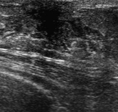
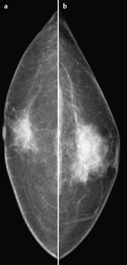
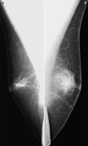
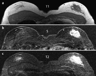
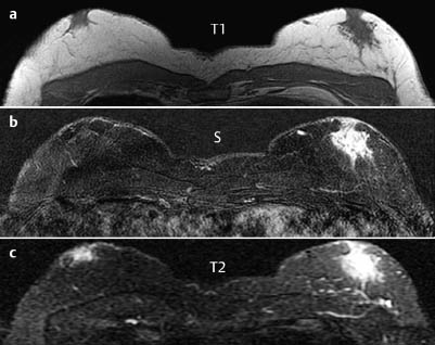
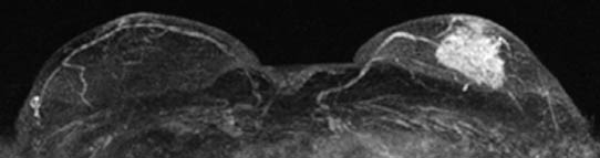
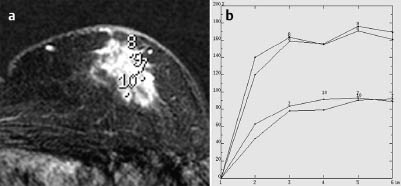
Case 39 Indication: Painful resistance in the left breast. History: Unremarkable. Risk profile: No increased risk. Age and sex: 62 years, male. Fig. 39.1 Ultrasound. Painful resistance 1 cm in diameter behind the areola of the left breast. Bilateral gynecomastia. Fig. 39.2a,b Digital mammography, CC view. Fig. 39.3a,b Digital mammography, MLO view. Fig. 39.4a–c Contrast-enhanced MR mammography. Fig. 39.5a–c Contrast-enhanced MR mammography. Fig. 39.6 Contrast-enhanced MR mammography. Maximum intensity projection. Fig. 39.7a,b Signal-to-time curves. Please characterize ultrasound, mammography, and MRI findings. What is your preliminary diagnosis? What are your next steps? These are the imaging studies of a symptomatic man. In the area of the resistance in the left breast, ultrasound showed ahypoechoic, lobulated mass (diameter 1 cm) with indeterminate distal echo pattern. US BI-RADS left 4. Mammograms demonstrated asymmetry of the parenchyma (left> right). There were no densities, architectural distortion, or microcalcifications. No circumscribed lesions. BI-RADS right 2/left 3.PGMI is not defined for imaging of men. MRI showed asymmetric parenchyma with partially ill-defined periphery. There was a notable unilateral enhancement of the left side after contrast administration, with a nonspecific signal curve. There were no unusual findings contralaterally. MRI Artifact Category: 1 MRI Density Type: 1
Clinical Findings

Ultrasound
Mammography
MR Mammography
MRM score | Finding right | Points |
Shape | irregular | 1 |
Border | ill-defined | 1 |
CM Distribution | inhomogenous | 1 |
Initial Signal Intensity Increase | strong | 2 |
Post-initial Signal Intensity Character | plateau | 1 |
MRI score (points) |
| 6 |
MRI BI-RADS |
| 5 |
 Preliminary Diagnosis
Right: Gynecomastia.
Left: Gynecomastia (asymmetrically to the other breast), mastitis, diffuse invasive carcinoma.
Clinical Findings | right 2 | left 3 |
Ultrasound | right 1 | left 4 |
Mammography | right 2 | left 3 |
MR Mammography | right 1 | left 5 |
BI-RADS Total | right 1 | left 5 |
Procedure
US-guided core biopsy.
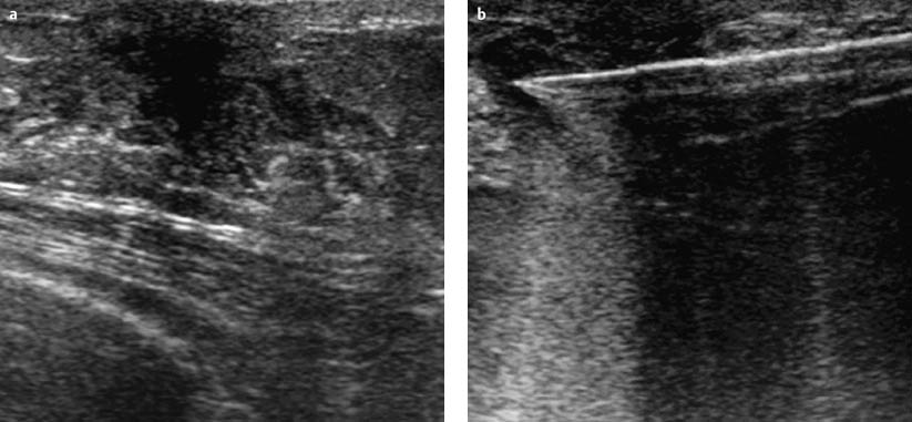
Fig. 39.8 a, b Pre-fire and post-fire documentation of US-guided core biopsy.
Histology
Adenosis.
Stay updated, free articles. Join our Telegram channel

Full access? Get Clinical Tree


