39 Collagenoses


10.1055/b-0034-77655

39 Collagenoses

Spindler-Thiele, S., Schmitt, R.

Clinically heterogeneous systemic diseases are included in the term collagenoses, which was coined by Klemperer in 1942. They have in common a generalized inflammatory reaction in the connective tissue with fibrinoid necroses and degeneration, as well as autoantibodies, which will not be further discussed here. Vasculitic diseases can be included in the classic collagenoses due to their clinical and immunological similarities. The following disease entities all affect the joints, and therefore illustrate the overlapping of symptoms within the systemic connective-tissue diseases.


Systemic Lupus Erythematosus (SLE)


Pathoanatomy and Clinical Symptoms

Antinuclear antibodies, among other pathophysiologic processes, lead to immune-complex vasculitis, which has an episodic course and primarily affects women of child-bearing age (female:male ratio = 10:1). A genetic predisposition is assumed because of the DR3 association in 60% of cases. The disease can be triggered by ultraviolet light, viruses, and hormonal changes. Lupus erythematosus is manifested in the skin (butterfly erythema), the musculoskeletal system, internal organs (nephritis, myocarditis, serositis), and the nervous system. In 90% of cases, there are arthralgias, which predominantly affect the joints of the hands and the knees with symmetric soft-tissue swelling.


Radiography

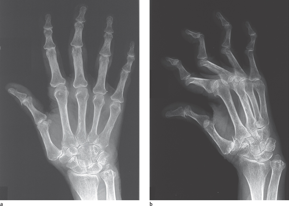
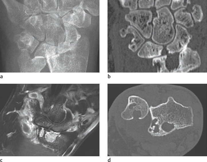
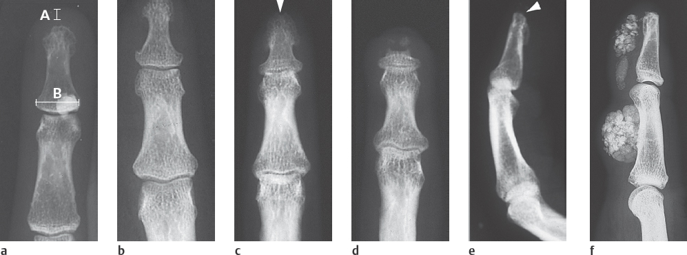
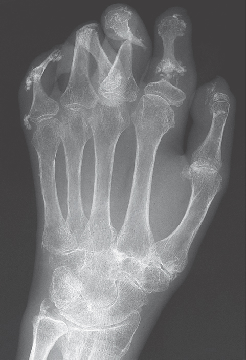
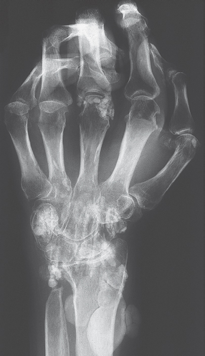
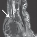
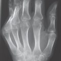
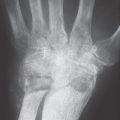
Typical findings are severe articular malalignments in the hands due to ligamentary instability without any other radiographic lesions ( Table 39.1 , Figs. 38.1c, 39.1 , 39.2 ).


Differential Diagnosis

Differential diagnoses include rheumatoid arthritis, which can be differentiated by the typical marginal erosions (see Figs. 36.1 and 38.1b ), Jaccoud arthropathy of rheumatic fever, which predominantly affect the ulnar side, Sharp syndrome, Ehlers-Danlos syndrome with articular malalignment, subcutaneous fat necroses, and signs of deforming osteoarthritis, as well as avascular osteonecroses.


Therapeutic Options

Depending on the disease activity, one can either wait or treat with immunosuppressive drugs. These include nonsteroidal anti-inflammatory drugs (NSAIDs), lowdose corticosteroids, hydroxychloroquine, azathioprine and even cyclophosphamide. The effectiveness of methotrexate, cyclosporine A, intravenous immunoglobulins, androgens, plasmapheresis, and immunoadsorption has not been confirmed.


Table 39.1 Radiographic signs of lupus erythematosus


  • Nonerosive, deforming arthropathies in about 10 % of cases:


    • Severe articular malalignment: Ulnar deviation of the metacarpophalangeal joints Subluxation of the trapeziometacarpal joint Elastic flexion and extension of the interphalangeal joints (swan-neck and button-hole deformities), may not appear in dorsopalmar radiographs because of positioning


    • Symmetric soft-tissue swelling


    • Periarticular osteopenia


  • Subcutaneous soft-tissue calcifications in fewer than 10% of cases


  • Ischemic osteonecroses in up to 3 %:


    • Epiphyseal osteonecroses usually in the metacarpal heads


    • Osteonecroses of the lunate and triquetrum


    • Cystoid radiolucency, increased density, fragmentation, and deformation of bones


  • Hooklike erosions are occasionally seen on the metaphyses


  • Acro-osteolysis and acro-osteosclerosis are rare


  • Raynaud phenomenon can be visualized in pharmacoangiography

Fig. 39.1a,b Arthropathic manifestation of systemic lupus erythematosus. Severe interphalangeal malalignment, which is well visualized in the semipronated oblique view. Swan-neck deformities with elastic hyperextension of the proximal interphalangeal joints and flexion in the distal interphalangeal joints. Subluxation of the trapeziometacarpal joint. No erosions. Periarticular osteopenia.
Fig. 39.2a-d Long-standing lupus erythematosus with articular instability and unusual erosions. a The dorsopalmar radiograph shows advanced destruction of the radiocarpal joint with ulnar translocation of the carpus. Large osteolyses in the radial epiphyse, and erosions in the trapezoid. b Coronal multiplanar reconstruction of an axial CT data set shows the extent of destruction on the radiocarpal joint compartment. Multiple carpal erosions and osteolyses. c Coronal T1-weighted SE sequence with fat saturation displays extensive contrast enhancing synovitis. d Axial CT scan shows a dislocation of the radius toward the palmar side. Note the deep erosions and osteolyses.


Scleroderma, Progressive Systemic Sclerosis (PSS)


Pathoanatomy and Clinical Symptoms

Overproduction of collagen and obliteration of small blood vessels leads to inflammatory, fibrotic, and regressive lesions in the cutis and subcutis, as well as in internal organs. Manifestations in the gastrointestinal tract, lungs, kidneys, heart, transverse-striated muscles, and bones result in the classic disease entity of progressive scleroderma, as well as a number of variants, which are listed in Table 39.2 . Painful micronecroses of the integument arise as a result of the underlying angiopathy. Immobile joints are a result of fibrosis of the synovium. Women in their third to fifth decades of life are primarily affected (female:male ratio = 3:1).


Table 39.2 Variants of scleroderma

Progressive systemic scleroderma (PSS)


Classic systemic manifestation at the skin and early visceral involvement


Thibièrge–Weissenbach syndrome


PSS combined with soft-tissue calcifications


CREST syndrome


Relatively benign variant of PSS:
C = calcinosis (Thibièrge–Weissenbach syndrome)
R = Raynaud phenomenon
E = esophageal dysfunction
S = sclerodactyly
T = telangiectasias


Sharp syndrome


Mixed connective-tissue disease (MCTD) is an overlapping syndrome of PSS with lupus erythematosus, polymyositis/dermatomyositis, and rheumatoid arthritis


Shulman syndrome


Eosinophilic fasciitis without involvement of internal organs, symmetric serous polyarthritis, rarely erosions, carpal tunnel syndrome


Focal scleroderma


Delineated sclerosis of the skin without involvement of internal organs


Secondary scleroderma


Caused by bleomycin, pentazocine, vinyl chloride, and solvents

Fig. 39.3a-f Radiographic appearance of acral lesions in scleroderma. a YUNE soft-tissue index for early detection of acral soft-tissue atrophy. Normally, AB/4. If AB/5, the condition is pathologic. Normal soft-tissue covering with areactive osteolysis on the unguinal tuberosity and soft-tissue calcinosis at the level of the distal interphalangeal joint. b Acral soft-tissue atrophy with “sugar-loaf” configuration in advanced scleroderma. c Early acro-osteolysis in the form of a so-called “rat-bite” defect (arrow). d Band-shaped acro-osteolysis. e Spotty acral soft-tissue sclerosis distal of the unguinal tuberosity. f Plaque-shaped calcinosis interstitialis localisata. Thibièrge-Weissenbach syndrome.
Fig. 39.4 Mutilation stage of scleroderma. Claw hand resulting from severe sclerodactyly. Destructive arthritis with areactive osteolyses in all phalanges and the ulnar styloid process. Nearly complete resorption of the third and fifth distal phalanges. Diffuse osteopenia. Interstitial calcinoses in the phalangeal soft tissues.
Fig. 39.5 Sharp syndrome (mixed connective-tissue disease). The flexion contractures of the fingers and the subluxations of metacarpophalangeal joints IV and V resemble lupus erythematosus; in contrast, the plaquelike and streaky soft-tissue calcifications are typical of dermatomyositis/polymyositis. Destruction of the ulnar styloid process and periarticular osteopenia. (Courtesy of A. Stäbler, MD, Munich.)


Table 39.3 Radiographic signs of scleroderma of the hand


  • Atrophy of soft tissues in 78 % of cases:


    • Sclerodactyly: atrophic skin of the fingers, which can lead to fixed claw hand, early diagnosis with the YUNE soft tissue index


    • Sugar-loaf finger form in advanced stages


  • Resorption of bone in up to 80 % of cases:


    • Areactive osteolyses on the fingertips, manifested early as “rat-bite” defects on the palmar side of the unguinal tuberosity


    • Resorption on trapeziometacarpal joint with radial subluxation of metacarpal I


    • Osteolyses on the radial and ulnar styloid processes


  • Soft-tissue calcifications in 25 % of cases:


    • Localized interstitial calcinosis: subcutaneous spotty sclerosis of the fingertips, also periarticular, rarely intra-articular


    • Universal interstitial calcinosis: diffuse calcifications, sometimes as large conglomerates


  • Osteoporosis in 80 % of cases:


    • Osteopenia of the hand skeleton – diffuse or band-shaped around joints


  • Destructive polyarthritis in about 25 % of cases:


    • Typically manifested on the proximal and distal interphalangeal joints without affecting the carpus or the metacarpophalangeal joints; radiocarpal and metacarpophalangeal involvement is rare


    • Destruction without bony proliferation; no cystic lesions


    • Mutilating arthritis is possible

Only gold members can continue reading. Log In or Register to continue

Stay updated, free articles. Join our Telegram channel

Jul 21, 2020 | Posted by in MUSCULOSKELETAL IMAGING | Comments Off on 39 Collagenoses

Full access? Get Clinical Tree

Get Clinical Tree app for offline access