5 Arteriography
A number of vascular variants are found in the forearm and hand, especially constitutional differences in the palmar arch resulting from a dominant radial or ulnar arterial supply. Indications for imaging of arteries in the forearm and hand are circulatory disturbances, inflammatory vascular diseases such as obliterating thrombangitis and traumatic vascular lesions, and the presurgical evaluation of congenital anomalies of the hand and vascularized soft-tissue tumors (especially hemangiomas). Catheter angiography with transfemoral or transbrachial accessneedle angiography is an invasive procedure. Advantages of these methods are the high spatial resolution with demonstration of even the smallest vessels, as well as functional recording of the blood flow. Contrast-enhanced magnetic resonance (MR) angiography differentiates between high-resolution and temporal-resolution techniques. Three-dimensional data sets can provide multiplanar maximal-intensity-projection (MIP) angiograms, whereas temporal-resolution data sets can only provide MIP images in the acquisition plane.
Anatomy and Variants of Hand Arteries
Forearm Arteries
The ulnar artery originates directly from the brachial artery. In the forearm, the arteria interossea communis, whose dorsal branch provides part of the arterial circulation of the back of the hand, branches off from it. In rare cases, there is a persisting median artery, which can join the superficial palmar arch. The ulnar artery divides into a superficial and a deep palmar arch in Guyon’s canal.
The radial artery originates in 80–85% of the cases from the brachial artery in the elbow. In the remaining 15–20%, it branches off high up from the brachial artery or the axillary artery, which can cause misinterpretation when the brachial artery is punctured. In theperiphery, the radial artery passes dorsally and crosses the trapezium and the base of the first metacarpal. In 96% of the cases, it runs along the palmar side of the hand in interdigital space I–rarely in II–or, after dividing into an accessory vessel, in interdigital spaces I and II. Next to the junction with the superficial and deep palmar arches, the main branches of the princeps pollicis artery and the radialis indicis artery, as well as the dorsal carpal network (to the rete carpi dorsalis) branch off from the radial artery in the periphery.

Arteries in the Palm
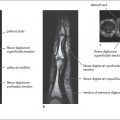
The palmar arteries represent a complex system of collaterals, which consists mainly of two palmar arches ( Fig. 5.1 ).
The superficial palmar arch is mainly fed by the ulnar artery. After passing through Guyon’s canal, the ulnar artery joins the superficial palmar arch, which ends superficially in relation to the flexor tendons of the fingers and the branches of the median nerve in the middle of the palm. The superficial palmar arch is closed in 42% of cases and anastomoses via its superficial radial branch with the radial artery. In 58% of cases, the arch is congenitally open. On the radial side of the hand, the common digital arteries originate from the superficial palmar arch, which projects onto the middle of the metacarpals in angiograms.
The deep palmar arch is fed primarily by the radial artery in 97% of the cases. Before reaching the deep palmar arch, however, the radial artery takes a dorsal detour by leaving the flexor side of the forearm at the level of the anatomical snuff box, and, after passing a short distance through metacarpal space I/II, returns to the palmar side. The deep palmar arch lies on top of the interosseous muscles and between the two heads of the adductor pollicis muscle deep in the middle of the palmar compartment. In 95% of cases, it is congenitally closed by an anastomosis with the deep network of the ulnar artery. In angiography, the deep palmar arch projects on the bases of the metacarpals, running proximal to the superficial arch. The princeps pollicis artery, radial indicis artery, and smaller palmar metacarpal arteries originate from the deep palmar arch.
Table 5.1 and Figure 5.2 summarize the arterial variants in the hand.
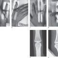
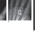
Figures 5.3 and 5.4 show examples of the arterial variations as depicted in angiograms.


Arteries of the Finger
In accordance with the wealth of variations in the palmar arch, the palmar metacarpal arteries, the common palmar digital arteries, and the properdigital arteries can have different sources of vascular supply ( Fig. 5.5 ). The most common vascular variants are summarized in Table 5.2 .

Related posts:


Stay updated, free articles. Join our Telegram channel

Full access? Get Clinical Tree


