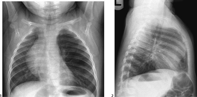
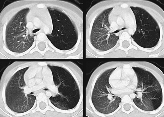
CASE 39 A 6-month-old presents with focal wheezing and mild respiratory distress. Figure 39A Frontal (Fig. 39A1) and lateral (Fig. 39A2) conventional chest radiographs demonstrate focal lucency and overinflation of the left upper lobe with mild shift of mediastinal structures to the right. Figure 39B Axial CT images of the child in Fig. 39A demonstrate left upper lobe lucency, attenuated vascularity, and overinflation, consistent with CLE. Congenital lobar emphysema (CLE) The definition of CLE is pathologic overdistention of a lobe on a congenital basis. It is one of the most common congenital pulmonary abnormalities. Despite its name, there is no true emphysema seen at pathology; that is, there is no alveolar wall destruction. Many have opted, therefore, to call this entity by the name of “congenital lobar overinflation.” CLE may occur secondary to a variety of causes. In approximately half of cases, no demonstrable cause can be found.
Clinical Presentation

Radiologic Findings

Diagnosis
Differential Diagnosis
Discussion
Background
Etiology
Stay updated, free articles. Join our Telegram channel

Full access? Get Clinical Tree


