CASE 39 A 48-year-old man presents with severe epigastric pain, nausea, vomiting, and diarrhea. Fig. 39.1 (A–F) Axial and coronal contrast-enhanced CT images show pancreatic calcification along with dilatation of the pancreatic duct. There is a multiloculated lesion seen in the anatomical plane between the pancreatic head and duodenum (pancreaticoduodenal groove). Postcholecystectomy clips are noted. Axial and coronal contrast-enhanced computed tomography (CT) images show pancreatic calcification along with dilatation of the pancreatic duct. There is a multiloculated lesion seen in the anatomical plane between the pancreatic head and the duodenum (pancreaticoduodenal groove). Postcholecystectomy clips are noted (Fig. 39.1). Groove pancreatitis Groove pancreatitis is a form of chronic segmental pancreatitis affecting the groove in the region of the pancreatic head, duodenum, and common bile duct. In 1982, Stolte et al coined the term groove pancreatitis
Clinical Presentation
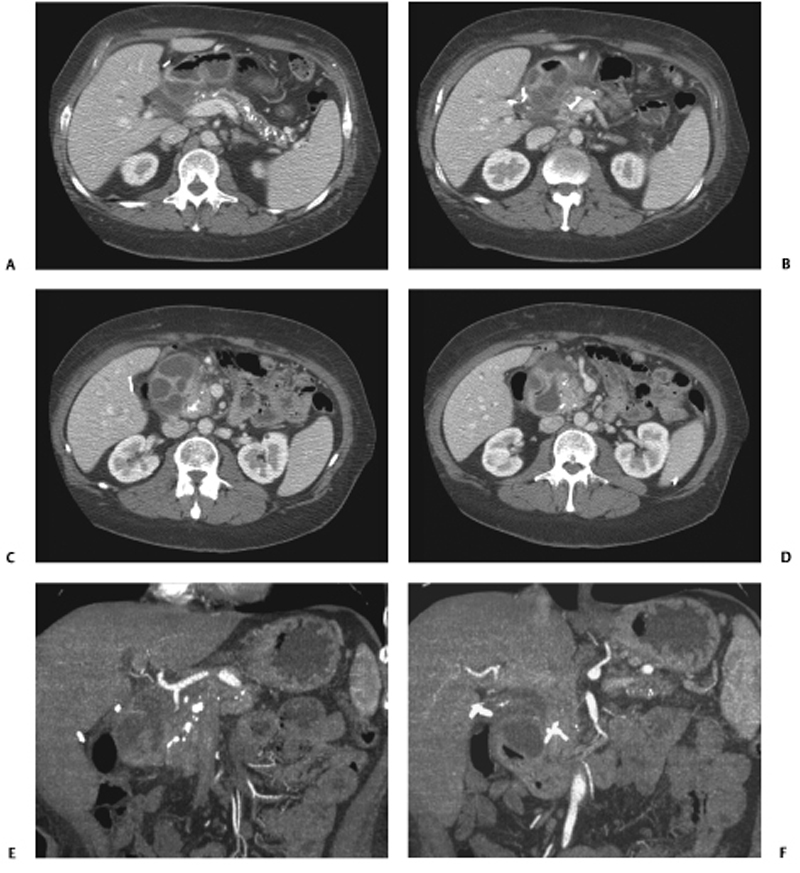
Radiologic Findings
Diagnosis
Differential Diagnosis
Discussion
Background
![]()
Stay updated, free articles. Join our Telegram channel


Full access? Get Clinical Tree


