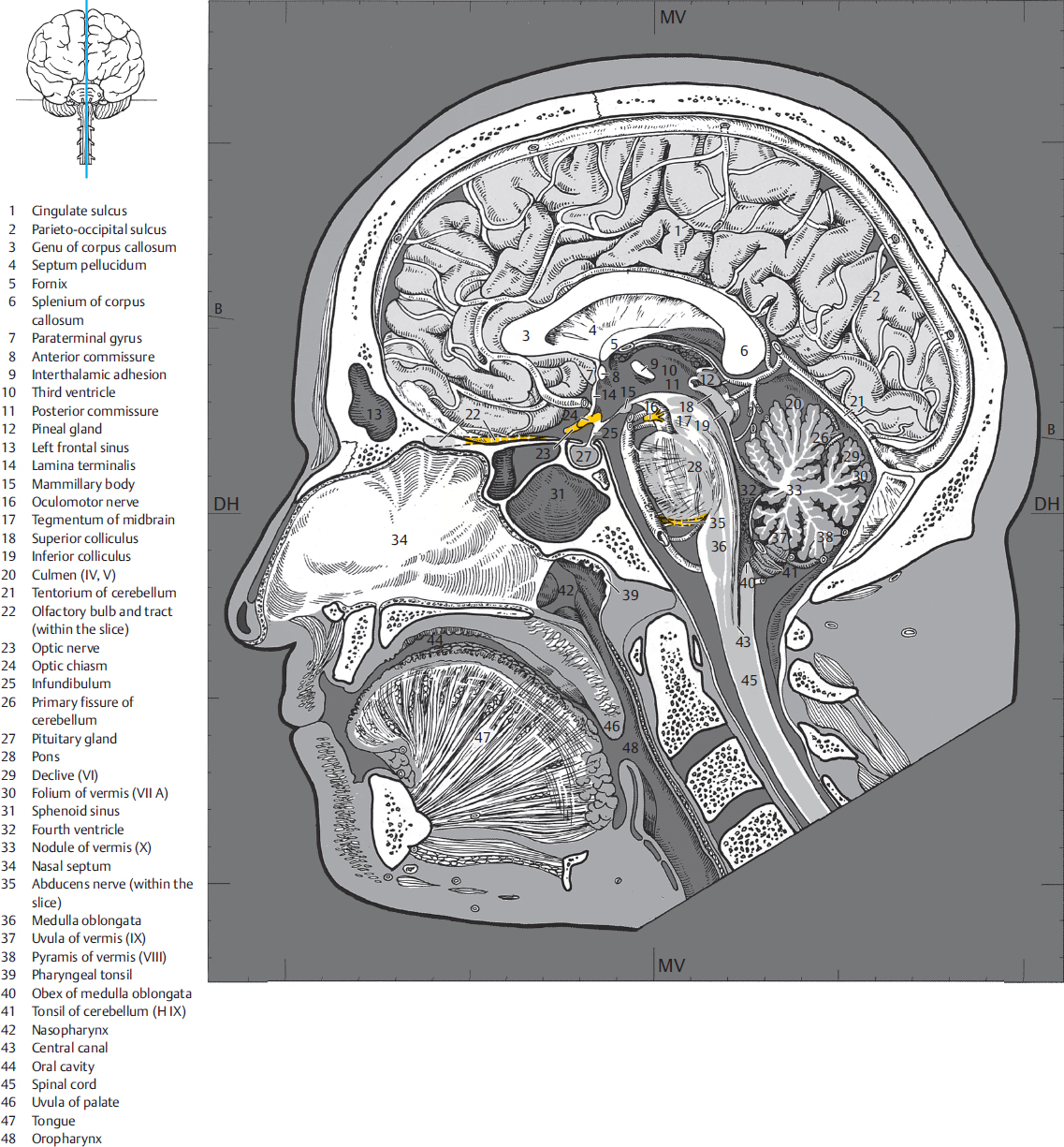
Sagittal MR images offer the advantage of the simultaneous depiction of facial bones and the cranial vault. The following structures are clearly seen in the median plane of the brain:
The craniocervical junction continues into the spinal cord. The typical shape of the corpus callosum and its lesions, as well as aplasia or atrophy thereof, are visualized in the median plane. The hypophysis and its pathological changes are also well delineated in the median plane and in a few paramedian sections. The bicommissural line is also well delineated in the median plane (see ▶Fig. 1.1), thereby enabling a transposition of brain anatomy in stereotactic–oriented atlases onto corresponding MR images using a coordinate system oriented in the bicommissural plane. Gyri and sulci of the endbrain are well defined in medial and lateral sagittal slices. Sulci run nearly perpendicular to the sectioned plane and are more distinctly visualized than the obliquely and tangentially coursing gyri due to the partial volume effect.
The ability to obtain sagittal sections counts as one of the merits of MRI, although sagittal reconstructions may also be obtained from CT volume data. Anomalies of midline structures of the brain are easily discernable on sagittal images; narrowing of the spinal canal by extradural space-occupying lesions or trauma sequelae may also be diagnosed (Section 9.2).
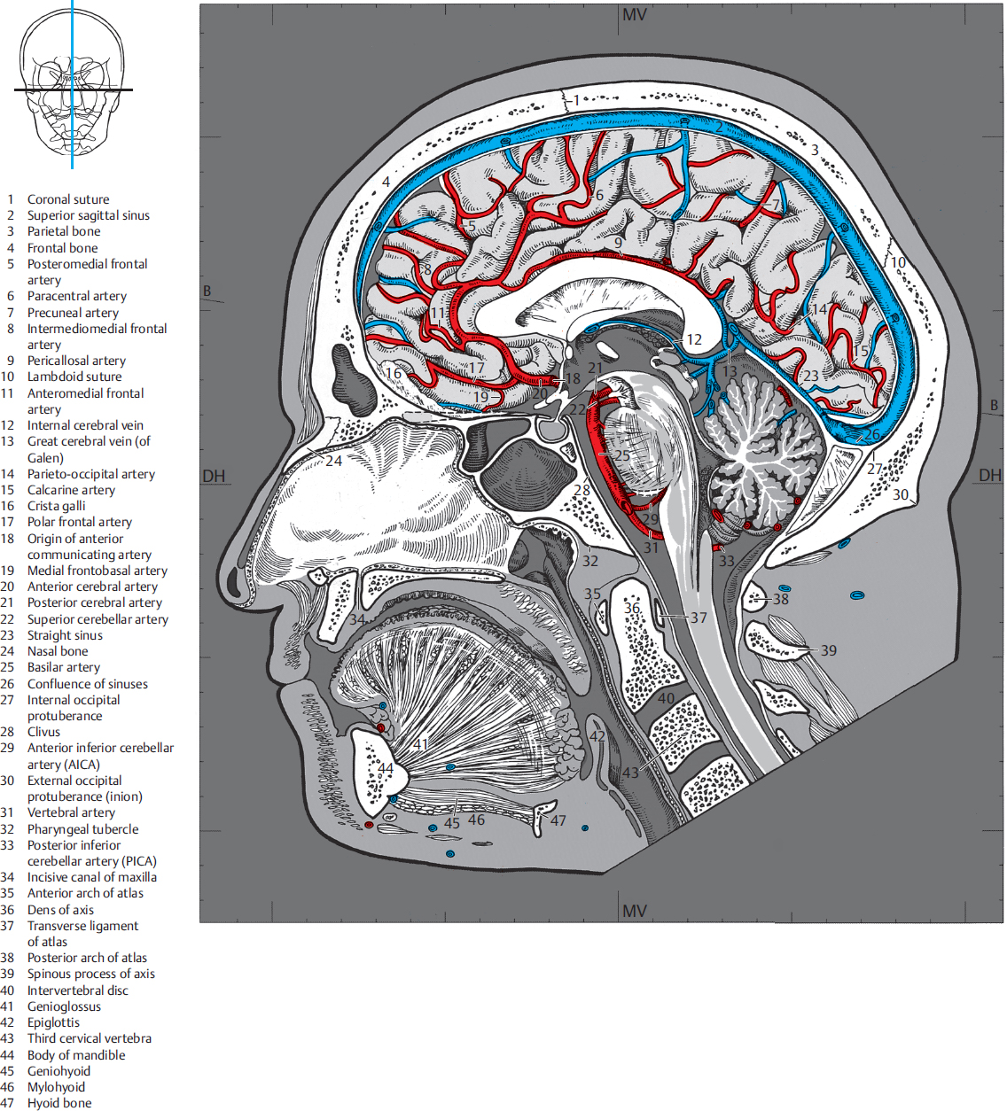
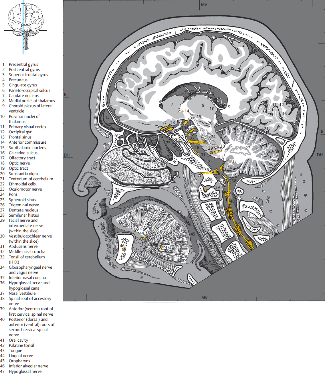
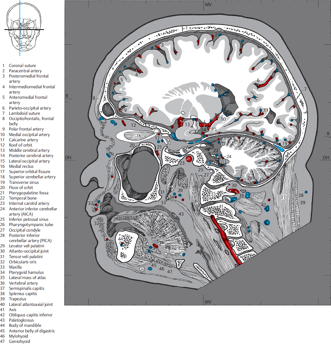
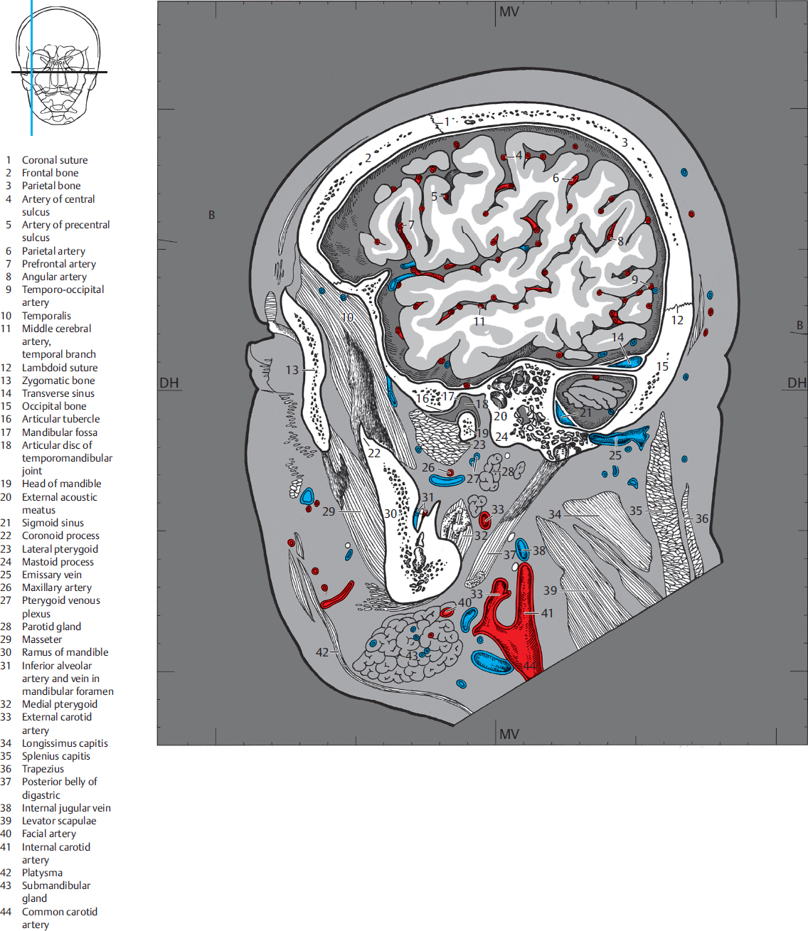
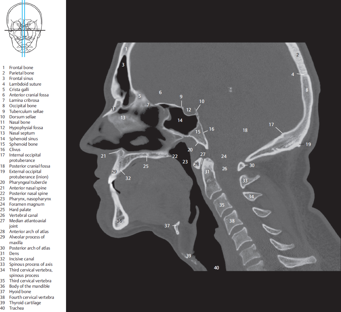
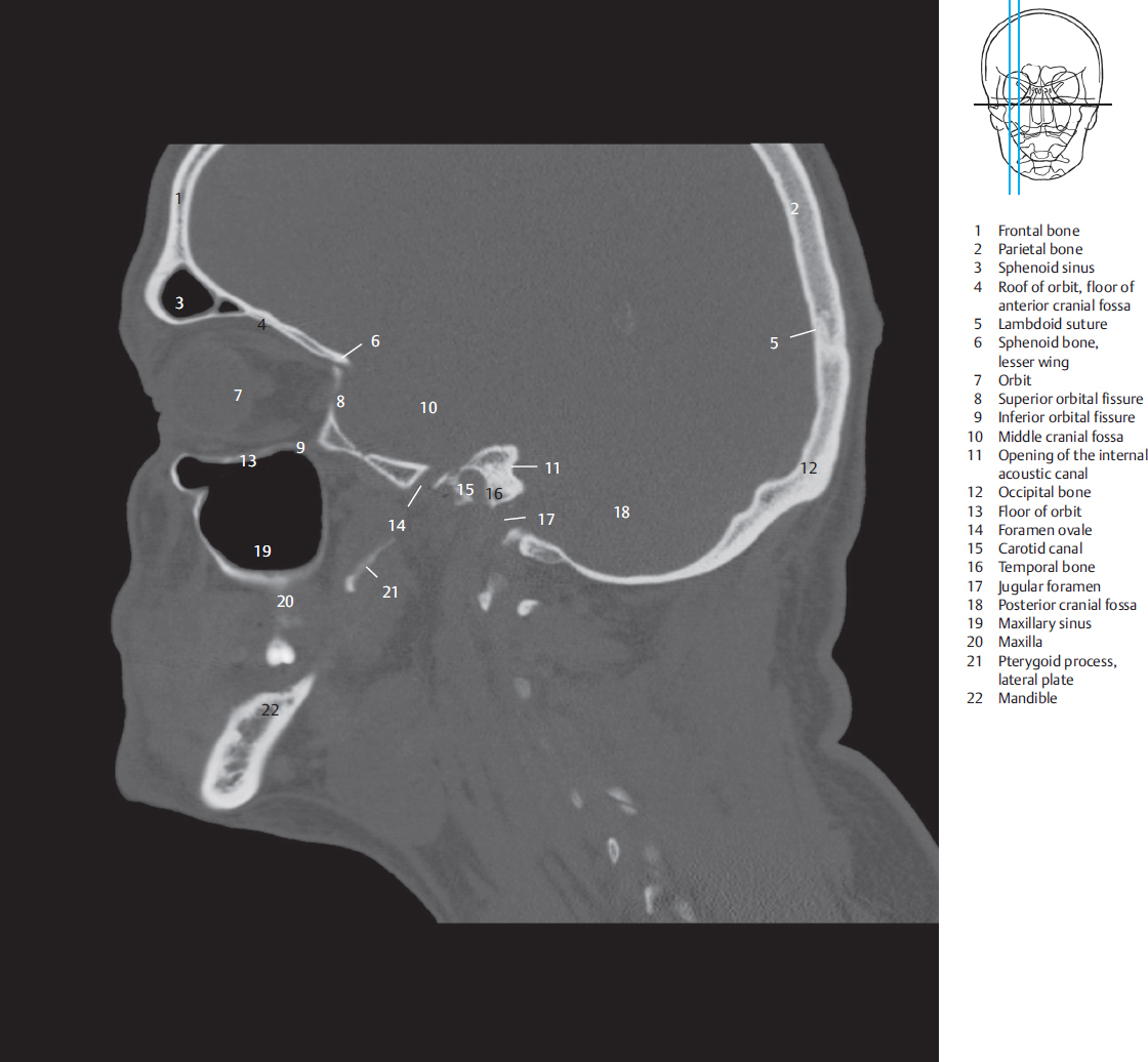
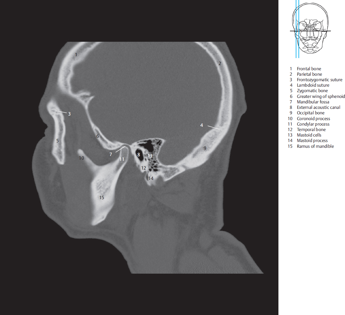
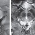
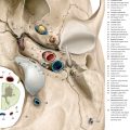
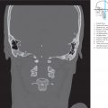
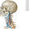
Fig. 4.1 Sagittal sections. The encircled digits indicate the number of 1-cm-thick sections. For details see ▶Chapter 12. DH = German horizontal.Fig. 4.1a Anterior view of the six sagittal sections. The illustrated cross-section always corresponds to the line lying medial to the encircled number of the slice concerned.Fig. 4.1b Illustration based on a cephalogram in an anteroposterior projection of the head from a. The six sagittal sections were drawn precisely and consecutively numbered from medial to lateral.Fig. 4.1c Anterior view of the brain in a and b. The coronal plane runs perpendicular to the German horizontal. Sagittal sections were assembled and numbered as in a.Fig. 4.1d View of the brain from a to c. Sagittal sections were assembled and numbered as in a.Fig. 4.2 Sagittal section.B = Bicommissural lineDH = German horizontalMV = Meatovertical lineFig. 4.2a Medial view of the first sagittal section. The falx cerebri has been removed, so that the medial surface of the endbrain is visible. The IIIrd ventricle, aqueduct and the IVth ventricle are recognizable as landmarks for the diencephalon and the brainstem. Nasal septum and paranasal sinuses together with the oral cavity, brain, and structures of the spinal cord. The Ist, IInd, IIIrd, and VIth cranial nerves have been graphically depicted, although they partly run within the slice.Fig. 4.2b Sagittal T1w MR image (T1w FLASH sequence), approximately corresponding to a. Both sagittal MR series (see ▶Fig. 4.2b, ▶Fig. 4.3b, ▶Fig. 4.4b, ▶Fig. 4.5b, ▶Fig. 4.6b, and ▶Fig. 4.7b, and ▶Fig. 4.2d, ▶Fig. 4.3d, ▶Fig. 4.4d, ▶Fig. 4.5d, ▶Fig. 4.6d, and ▶Fig. 4.7d were obtained from a 33-year-old man (volunteer). For technical data, see ▶Chapter 12.Fig. 4.2c Medial view of the first sagittal section of the right half of the head. The cranial cavity was opened in the plane of the crista galli and the internal occipital protuberance. The spinal canal has been halved in the midline at the level of the upper cervical vertebrae. Bony structures, muscles, and blood vessels.Fig. 4.2d Sagittal T2w MR image, approximately corresponding to c.Fig. 4.3 Sagittal section.B = Bicommissural lineDH = German horizontalMV = Meatovertical lineFig. 4.3a Medial view of the second sagittal section. The anterior and central parts of the right lateral ventricle are visible. The lateral aspects of the midbrain and the pons are the only parts of the brainstem that have been sectioned in this slice. The spinal roots of the XIth cranial nerve and Ist spinal nerve can be identified in the vertebral canal. The enlarged turbinates have been tangentially sectioned in the nasal cavity. Paranasal sinuses, oral cavity, brain structures, as well as cranial and spinal nerves.Fig. 4.3b Sagittal T1w MR image, approximately corresponding to the sectional plane in a and c. For technical data see ▶Chapter 12.Fig. 4.3c Medial view of the second sagittal section. The section lying at a distance of 1 cm from the median plane lies lateral to the pituitary fossa and runs through the cavernous sinus and the foramen magnum. The orbit lies lateral to the section and has therefore not been opened. Bony structures, muscles, and blood vessels.Fig. 4.3d Sagittal T2w MR image, approximately corresponding to the sectional plane in a and c. For technical data see ▶Chapter 12.Fig. 4.4 Sagittal section.B = Bicommissural lineDH = German horizontalMV = Meatovertical lineFig. 4.4a Medial view of the third sagittal section. The section lies lateral to the brainstem at the level of the medial geniculate body. The temporal lobe has been sectioned tangentially. Paranasal sinuses, oral cavity, brain structures, cranial nerves, and the first spinal nerves.Fig. 4.4b Sagittal T1w MR image, approximately correspon ding to the sectional plane in a and c.Fig. 4.4c Medial view of the third sagittal section. The section runs through the orbital apex, medial aspect of the superior orbital fissure, middle cranial fossa, and the parapharyngeal space. Bony structures, muscles, and blood vessels.Fig. 4.4d Sagittal T2w MR image, approximately corresponding to the sectional plane in a and c.Fig. 4.5 Sagittal section.B = Bicommissural lineDH = German horizontalMV = Meatovertical lineFig. 4.5a Medial view of the fourth sagittal section. Parts of the endbrain lying lateral to the anterior horn and cella media of the lateral ventricle are visible in the supratentorial region, as also those which abut the inferior horn, like the hippocampus and the amygdala. The section runs through the cerebellar hemispheres in the infratentorial region. Brain structures and cranial nerves.Fig. 4.5b Sagittal T1w MR image, approximately corresponding to the sectional plane in a and c.Fig. 4.5c Medial view of the fourth sagittal section. The section is closely apposed medially to the midplane of the eyeball, thereby sectioning the lens of the eye and the superior and inferior recti. The internal auditory canal, jugular foramen, and the parapharyngeal space are identifiable in this section. Bony structures, muscles, and blood vessels.Fig. 4.5d Sagittal T2w MR image, approximately corresponding to the sectional plane in a and c.Fig. 4.6 Sagittal section.B = Bicommissural lineDH = German horizontalMV = Meatovertical lineFig. 4.6a Medial view of the fifth sagittal section. The insula has been tangentially sectioned and lies in the lateral sulcus, surrounded by the insular arteries. Branches of the Vth, VIIth, Xth, XIth, and XIIth cranial nerves are visible. Brain structures and cranial nerves.Fig. 4.6b Sagittal T1w MR image, approximately corresponding to the sectional plane in a and c.Fig. 4.6c Medial view of the fifth sagittal section. The lateral part of the eyeball has been sectioned. The skull base has been sectioned at the level of the cochlea. Bony structures, muscles, and blood vessels.Fig. 4.6d Sagittal T2w MR image, approximately corresponding to the sectional plane in a and c.Fig. 4.7 Sagittal section.B = Bicommissural lineDH = German horizontalMV = Meatovertical lineFig. 4.7a Medial view of the sixth sagittal section. Parts of the cortex of the endbrain have been evenly sectioned, especially those which surround the lateral sulcus as operculum. Brain structures and cranial nerves.Fig. 4.7b Sagittal T1w MR image, approximately corresponding to the sectional plane in a and c.Fig. 4.7c Medial view of the sixth sagittal section. This section lies close to the orbit at its lateral aspect and contains the bony part of the auditory canal and the carotid bifurcation. Bony structures, muscles, and blood vessels.Fig. 4.7d Sagittal T2w MR image, approximately corresponding to the sectional plane in a and c.Fig. 4.8 Sagittal section. Sagittal CT image, the position of which approximately corresponds to that of the first sagittal section. This and the following sagittal CT images were obtained during a diagnostic examination; the cranial vault is therefore incompletely visualized. Secondary sagittal reconstruction from a thin, reconstruction-enabling transverse data set. Landmarks are the pituitary fossa, frontal and sphenoid sinuses, atlas, and dens.Fig. 4.9 Sagittal section. CT image, the position of which approximately corresponds to that of the second sagittal section. The floor of the anterior and posterior cranial fossae has been sectioned in the cranial cavity. Landmarks are the ethmoidal cells, hard palate, mandible, and the contours of cervical vertebrae.Fig. 4.10 Sagittal section. CT image, the position of which approximately corresponds to that of the third sagittal section. The roof and floor of orbit, maxillary sinus, mandible, and the contours of the cervical vertebrae are landmarks for the facial skeleton and the craniocervical junction.Fig. 4.11 Sagittal section. Sagittal CT image, corresponding to the fourth sagittal section. The terraces of the anterior, middle, and posterior cranial fossae are clearly delineated.Fig. 4.12 Sagittal section. CT image, corresponding to the fifth sagittal section. The skull base has been sectioned at the level of the superior semicircular canal, the facial canal, the tympanic cavity, and the styloid process.Fig. 4.13 Sagittal section. CT image, corresponding to the sixth sagittal section. The section lies at the level of the temporomandibular joint and contains the mastoid process.
Only gold members can continue reading. Log In or Register to continue