10.1055/b-0039-166440

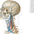
Fig. 3.1 Coronal sections. Encircled digits indicate the number of 1-cm-thick sections as viewed from the front. For details see ▶Chapter 12. Roman numbers in the legends refer to the numerical identifiers of parts of the cerebellum (see ▶Table 7.1). DH = German horizontal. Fig. 3.1a Position of coronal sections in lateral view. The portrayed cross-section always corresponds to the line lying anterior (in the image on the left) to the encircled number of the slice concerned. 
Fig. 3.1b Drawing based on a cephalogram from a. Cross-sections of 14 coronal sections were optimally transposed on to an X-ray image and numbered consecutively from anterior to posterior. 
Fig. 3.1c Median view of the brain and upper spinal cord of the same head as in a and b. Coronal sections were assembled and numbered as in a. 
Fig. 3.1d Lateral view of the brain and the upper spinal cord of the same head as in a to c. Coronal sections were assembled and numbered as in a. 
Fig. 3.2 1st coronal section. DH = German horizontal M = median plane Fig. 3.2a Anterior view of first coronal section. The blue line on the top left shows the course of the sectional plane through the anterior part of the frontal lobe (see ▶Fig. 3.1c). Nasal cavity, paranasal sinuses, oral cavity, brain structures, retina, and cranial nerves. 
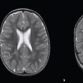
Fig. 3.2b Coronal T1w MR image (T1w FLASH sequence), corresponding approximately to the sectional plane in a and c. Brain structures are accentuated on the selected sequence with a field strength (magnetic flux density) of 3T. The two coronal MR series (see ▶Fig. 3.2b, ▶Fig. 3.3b, ▶Fig. 3.4b, ▶Fig. 3.5b, ▶Fig. 3.6b, ▶Fig. 3.7b, ▶Fig. 3.8b, ▶Fig. 3.9b, ▶Fig. 3.10b, ▶Fig. 3.11b, ▶Fig. 3.12b, ▶Fig. 3.13b, ▶Fig. 3.14b, ▶Fig. 3.15b, ▶Fig. 3.2d, ▶Fig. 3.3d, ▶Fig. 3.4d, ▶Fig. 3.5d, ▶Fig. 3.6d, ▶Fig. 3.7d, ▶Fig. 3.8d, ▶Fig. 3.9d, ▶Fig. 3.10d, ▶Fig. 3.11d, ▶Fig. 3.12d, ▶Fig. 3.13d, ▶Fig. 3.14d, and ▶Fig. 3.15d) were obtained from a 33-year-old man. 
Fig. 3.2c Anterior view of the first coronal section. The blue line on the top left marks the position of the sectional plane at the level of the crista galli and the orbit (see ▶Fig. 3.1b). Bony structures, muscles, and blood vessels. 
Fig. 3.2d Coronal T2w MR image, corresponding approximately to the sectional plane in a and c. 
Fig. 3.3 2nd coronal section. DH = German horizontal M = median plane Fig. 3.3a Anterior view of the 2nd coronal section. The frontal lobe has been sectioned at the level of the olfactory bulb. Nasal cavity, paranasal sinuses, and cranial nerves. 
Fig. 3.3b Coronal T1w MR image, corresponding approximately to the sectional plane in a and c. 
Fig. 3.3c Anterior view of the 2nd coronal section. The section lies approximately 6 mm behind the eyeball and roughly in the center of the body of the mandible. Bony structures, muscles, and blood vessels. 
Fig. 3.3d Coronal T2w MR image, corresponding approximately to the sectional plane in a and c. 
Fig. 3.4 3rd coronal section. DH = German horizontal M = median plane Fig. 3.4a Anterior view of the 3rd coronal section. The sectional plane lies in the frontal lobe approximately 8 mm anterior to the genu of corpus callosum (see ▶Fig. 3.1c). The optic nerve, branches of the trigeminal nerve, and the hypoglossal nerve have been sectioned in the region of the facial skeleton. Nasal cavity, paranasal sinuses, oral cavity, brain structures, and cranial nerves. 
Fig. 3.4b Coronal T1w MR image, corresponding approximately to the sectional plane in a and c. 
Fig. 3.4c Anterior view of the 3rd coronal section. The sectional plane runs close behind the center of the anterior cranial fossa, through the posterior third of the orbit and the coronoid process of the mandible. Bony structures, muscles, and blood vessels. 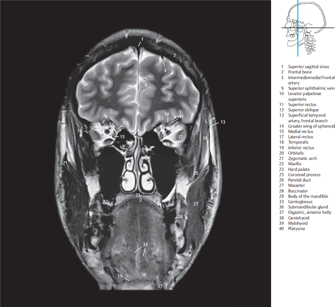
Fig. 3.4d Coronal T2w MR image, corresponding approximately to the sectional plane in a and c. 
Fig. 3.5 4th coronal section. DH = German horizontal M = median plane Fig. 3.5a Anterior view of the 4th coronal section. The frontal lobe has been sectioned at the level of the genu of corpus callosum. The abducent, oculomotor, and trochlear nerves, and the optic nerve lie close to each other at the orbital apex. The hypoglossal nerve and branches of the mandibular nerve lie in the region of the floor of the mouth. Nasal cavity and paranasal sinuses, brain structures, and cranial nerves. 
Fig. 3.5b Coronal T1w MR image. 
Fig. 3.5c Anterior view of the 4th coronal section. The anterior poles of the middle cranial fossa have been sectioned and lie under the lesser wings of the sphenoid. The optic canal and the superior orbital fissure can be identified in the sectional plane. The tongue has been sectioned at the level of the hyoid bone. Bony structures, muscles, and blood vessels. 
Fig. 3.5d Coronal T2w MR image. 
Fig. 3.6 5th coronal section. DH = German horizontal M = median plane Fig. 3.6a Anterior view of the 5th coronal section. The frontal horns of the lateral ventricles have been sectioned in this plane. The optic chiasma lies in this slice. The lingual and hypoglossal nerves are identified lateral to the pharyngeal isthmus. Brain structures and cranial nerves. 
Fig. 3.6b Coronal T1w MR image, corresponding to the sectional plane in a and c. 
Fig. 3.6c Anterior view of the 5th coronal section. The sectional plane lies approximately in the center of the sphenoid bone at the level of the anterior clinoid process and in the center of the zygomatic arch. The nasopharynx, soft palate, and the uvula are visualized in this section. Bony structures, muscles, an blood vessels. 
Fig. 3.6d Coronal T2w MR image, corresponding to the sectional plane in a and c. 
Fig. 3.7 6th coronal section. DH = German horizontal M = median plane Fig. 3.7a Anterior view of the 6th coronal section. The sectional plane lies in the median region 2 mm anterior to the anterior commissure (interrupted lines within the slice). Its anterior extensions section the basal ganglia laterally. The abducens, oculomotor, and trochlear nerves lie lateral to the hypophysis. Brain structures and cranial nerves. 
Fig. 3.7b Coronal T1w MR image, which corresponds to the plane in a and c. 
Fig. 3.7c Anterior view of the 6th coronal section. The sectional plane runs through the pituitary fossa at the level of the posterior clinoid processes and through both temporomandibular joints. The posterior wall of the pharynx lies in the anterior half of the 1-cm-thick slice. Bony structures, muscles, and blood vessels. 
Fig. 3.7d Coronal T2w MR image, corresponding to the section in a and c. 
Fig. 3.8 7th coronal section. DH = German horizontal M = median plane Fig. 3.8a Anterior view of the 7th coronal section. The interventricular foramina, the mammillary bodies and the anterior part of the inferior horn of the lateral ventricle lie in this plane. Brain structures and cranial nerves. 
Fig. 3.8b Coronal T1w MR image, corresponding to the section in a and c. 
Fig. 3.8c Anterior view of the 7th coronal section. The sectional plane lies at the level of the anterior wall of the cartilaginous external auditory canal. Vertebral bodies have been evenly sectioned. The internal carotid arteries are seen ascending within this slice. Bony structures, muscles, and blood essels. 
Fig. 3.8d Coronal T2w MR image, corresponding to the section in a and c. 
Fig. 3.8e Coronal schematic drawing of the superior sagittal sinus, displaying its structure with arachnoid granulations. (Reproduced from Schuenke, Schulte, and Schumacher, Atlas of Anatomy, 2nd edition, ©2014, Thieme Publishers, New York. Illustration by Karl Wesker/Markus Voll.) 
Fig. 3.9 8th coronal section. DH = German horizontal M = median plane Fig. 3.9a Anterior view of the 8th coronal section. The sectional plane runs through the endbrain, interbrain, midbrain and pons at the level of the interpeduncular fossa. The IVth, Vth, VIth, VIIth, and VIIIth cranial nerves are visualized within the cranial cavity while the VIIth, IXth, XIth, and XIIth nerves are seen outside of it. Brain structures and cranial nerves. 
Fig. 3.9b Coronal T1w MR image, approximately corresponding to the sectional plane in a and c. 
Fig. 3.9c Anterior view of the 8th coronal section. The bony external auditory canal, tympanic cavity and the internal auditory canal lie in this slice. The internal jugular veins course through the slice lateral to the dens and the cervical vertebral bodies. Bony structures, muscles, and blood vessels. 
Fig. 3.9d Coronal T2 w MR image, approximately corresponding to the sectional plane in a and c. 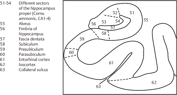
Fig. 3.9e Depiction of the anatomical structure of the hippocampus. (Source: [480]) 
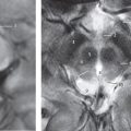
Fig. 3.9f High resolution T2w MR image of the hippocampal formation with a field strength of 7T. (Source: With kind permission of Dr. J. Theysohn, University Hospital Essen.) 
Fig. 3.10 9th coronal section. DH = German horizontal M = median plane Fig. 3.10a Anterior view of the 9th coronal section. The posterior commissure has been sectioned in this slice. The lateral ventricle with cella media and inferior horn as well as the first part of the cerebral aqueduct can be seen. The VIIth, VIIIth, IXth, Xth, XIth, and XIIth cranial nerves arise from the brainstem. The quadrigeminal plate lies within the slice and is therefore not visible (see ▶Fig. 3.1c). 
Fig. 3.10b Coronal T1w MR image, approximately corresponding to the sectional plane in a and c. 
Fig. 3.10c Anterior view of the 9th coronal section. The slice lies approximately in the center of the mastoid process, opening up the posterior cranial fossa and the upper spinal canal in the region of the first four cervical vertebrae. Bony structures, muscles, and blood vessels. 
Fig. 3.10d Coronal T2w MR image, approximately corresponding to the sectional plane in a and c. 
Fig. 3.11 10th coronal section. DH = German horizontal M = median plane Fig. 3.11a Anterior view of the 10th coronal section. The splenium of the corpus callosum, collateral trigone of the lateral ventricle, the pineal gland, the floor of the rhomboid fossa, and the cerebellum as the floor of the IVth ventricle have been sectioned in the slice. Brain structures and branches of spinal nerves. 
Fig. 3.11b Coronal T1w MR image, approximately corresponding to the sectional plane in a and c. 
Fig. 3.11c Anterior view of the 10th coronal section. The section lies just behind the petrous part of the temporal bone and runs directly behind the center of the foramen magnum (see ▶Fig. 3.1a). The upper cervical vertebrae have been sectioned at the level of the vertebral arch. Bony structures, muscles, and blood vessels. 
Fig. 3.11d Coronal T2w MR image, approximately corresponding to the sectional plane in a and c. 
Fig. 3.12 11th coronal section. DH = German horizontal M = median plane Fig. 3.12a Anterior view of the 11th coronal section. The posterior horn of the lateral ventricle forms a landmark in the supratentorial region. The cerebellum with the dentate nucleus is located in the infratentorial region. Brain structures and branches of spinal nerves. 
Fig. 3.12b Coronal T1w MR image, approximately corresponding to the sectional plane in a and c. 
Fig. 3.12c Anterior view of the 11th coronal section. The posterior part of the foramen magnum lies in the slice. Bony structures, neck muscles, and blood vessels. 
Fig. 3.12d Coronal T2w MR image, approximately corresponding to the sectional plane in a and c. 
Fig. 3.13 12th coronal section. DH = German horizontal M = median plane Fig. 3.13a Anterior view of the 12th coronal section. The posterior horn of the lateral ventricle has only been sectioned in the left cerebral hemisphere. The tentorium of cerebellum completely separates the supratentorial and infratentorial regions. Brain structures and branches of spinal nerves. 
Fig. 3.13b Coronal T1w MR image, approximately corresponding to the sectional plane in a and c. 
Fig. 3.13c Anterior view of the 12th coronal section. The slice lies behind the foramen magnum. Only three spinous processes of cervical vertebrae have been sectioned. Bony structures, neck muscles, and blood vessels. 
Fig. 3.13d Coronal T2w MR image, approximately corresponding to the sectional plane in a and c. 
Fig. 3.14 13th coronal section. DH = German horizontal M = median plane Fig. 3.14a Anterior view of the 13th coronal section. Only parts of the parietal and occipital lobes have been sectioned in the endbrain. Tangential section through the cerebellum. Brain structures and branches of spinal nerves. 
Fig. 3.14b Coronal T1w MR image, approximately corresponding to the sectional plane in a and c. 
Fig. 3.14c Anterior view of the 13th coronal section. The parietal and occipital bones form a rounded bony ring. Bony structures, neck muscles, and blood vessels. 
Fig. 3.14d Coronal T2w MR image, approximately corresponding to the sectional plane in a and c. 
Fig. 3.15 14th coronal section. DH = German horizontal M = median plane Fig. 3.15a Anterior view of the 14th coronal section. The posterior part of both cerebral hemispheres is composed almost entirely of the occipital lobes. Brain structures and branch of the second spinal nerve. 
Fig. 3.15b Coronal T1w MR image, approximately corresponding to the sectional plane in a and c. 
Fig. 3.15c Anterior view of the 14th coronal section, which contains the confluence of sinuses. The curvature of the neck created a gap while sectioning. Bony structures, neck muscles, and blood vessels. 
Fig. 3.15d Coronal T2w MR image, approximately corresponding to the sectional plane in a and c. 
Fig. 3.16 1st coronal section. CT image, the position of which approximately corresponds to that of the first coronal section. This and the following CT images were obtained during a diagnostic examination; the calvarium is therefore incompletely depicted. Secondary coronal reconstruction from a thin, reconstruction-enabling transverse data set. Landmarks are the anterior cranial fossa with crista galli, the orbits, ethmoid cells, nasal turbinates, maxillary sinuses, and the mandible. 
Fig. 3.17 2nd coronal section. CT image, the position of which approximately corresponds to that of the second coronal section. Bony landmarks are the roof and floor of the orbit, nasal turbinates, maxillary sinuses, and the mandible. 
Fig. 3.18 3rd coronal section. CT image, the position of which approximately corresponds to that of the 4th coronal section. The illustration depicts the sphenoid bone, pterygopalatine fossa, and the optic canal. 
Fig. 3.19 4th coronal section. CT image, the position of which approximately corresponds to that of the 5th coronal section and includes the foramen rotundum and the pterygoid process of the sphenoid bone. The central part of the hyoid bone is also visible. 
Fig. 3.20 5th coronal section. CT image, the position of which approximately corresponds to that of the 6th coronal section. The middle cranial fossa has been sectioned at the level of the posterior clinoid processes, sphenoid sinus, ramus of the mandible, and the lateral parts of the hyoid bone. 
Fig. 3.21 6th coronal section. CT image, the position of which approximately corresponds to that of the 7th coronal section. The middle cranial fossa has been sectioned at the level of the temporomandibular joints, the lateral parts of the hyoid bone, and the anterior arch of the first cervical vertebra. 
Fig. 3.22 7th coronal section. CT image, the position of which approximately corresponds to that of the 8th coronal section. It includes the petrous part of the temporal bone with the cochlea and the styloid process as well as the anterior part of the upper cervical spine. 
Fig. 3.23 8th coronal section. CT image, mildly posteriorly positioned than the 8th coronal section. The internal auditory canal, facial canal, and mastoid air cells are visible in the petrous part of the temporal bone. The first three cervical vertebrae are well seen. 
Fig. 3.24 9th coronal section. CT image, the position of which approximately corresponds to that of the 9th coronal section. The posterior cranial fossa, which transitions to the spinal canal at the foramen magnum, lies medial to the petrous parts of the temporal bones. 
Fig. 3.25 10th coronal section. CT image, the position of which approximately corresponds to that of the 10th coronal section. The calvarium forms a bony ring (incompletely seen here) with a caudal opening at the foramen magnum.
3 Coronal Sections
Fourteen coronal sections have been described in this part of the book from anterior to posterior. ▶Fig. 3.1 describes the position of coronal sections; ▶Fig. 3.2, ▶Fig. 3.3, ▶Fig. 3.4, ▶Fig. 3.5, ▶Fig. 3.6, ▶Fig. 3.7, ▶Fig. 3.8, ▶Fig. 3.9, ▶Fig. 3.10, ▶Fig. 3.11, ▶Fig. 3.12, ▶Fig. 3.13, ▶Fig. 3.14, ▶Fig. 3.15, ▶Fig. 3.16, ▶Fig. 3.17, ▶Fig. 3.18, ▶Fig. 3.19, ▶Fig. 3.20, ▶Fig. 3.21, ▶Fig. 3.22, ▶Fig. 3.23, ▶Fig. 3.24, and ▶Fig. 3.25 reproduce anterior views of coronal sections.
Related posts:


Stay updated, free articles. Join our Telegram channel

Full access? Get Clinical Tree


