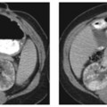
CASE 40 An 81-year-old man presents with a history of bloating and epigastric pain. Fig. 40.1 (A–C) Axial contrast-enhanced images in the pancreatic phase of CT shows a large enhancing lesion in the head of the pancreas (arrow). There was no intrahepatic ductal dilatation. The portal vein and the superior mesenteric artery were patent and not involved. Axial contrast-enhanced images in the pancreatic phase of computed tomography (CT) show a large enhancing lesion in the head of the pancreas (Fig. 40.1). There was no intrahepatic ductal dilatation. The portal vein and the superior mesenteric artery were patent and not involved. Acinar cell carcinoma of the pancreas
Clinical Presentation

Radiologic Findings
Diagnosis
Differential Diagnosis
Discussion
Background
Stay updated, free articles. Join our Telegram channel

Full access? Get Clinical Tree


