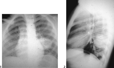
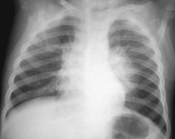
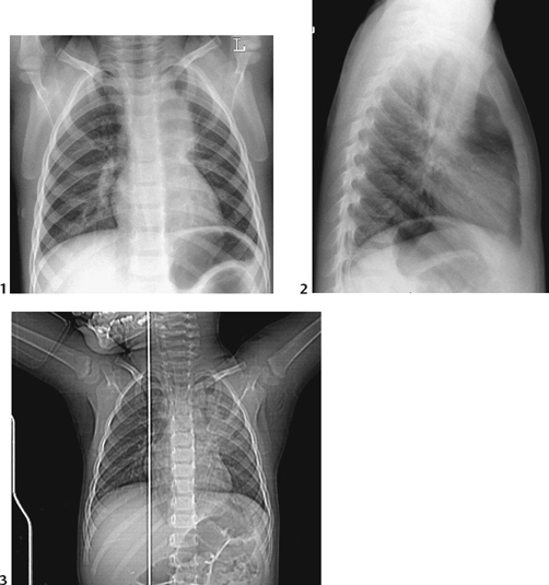
CASE 40 A previously well 4-year-old presents with acute onset cough, leukocytosis, and fever. Figure 40A The AP chest radiograph shows spherical mass-like areas of soft tissue in the right upper lobe of the lung, which is sharply defined and with uniform opacity consistent with typical “round pneumonias” (Fig. 40A). Figure 40B (1,2) Standard chest radiographs of a child with cough and fever showing well-defined and sharply marginated areas of soft tissue density within the lung, consistent with typical round pneumonias. Figure 40C Large, sharply marginated left perihilar soft tissue masslike opacity in the region of the superior segment of the left lower lobe in a child with cough and fever. Round pneumonia Round pneumonias are most commonly seen in children ≤8 years of age. They are a unique radiographic manifestation of a usual bacterial pneumonia in this age range.
Clinical Presentation
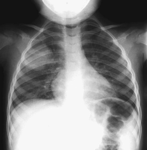
Radiologic Findings
Diagnosis
Differential Diagnosis
Discussion
Background
Related posts:


Stay updated, free articles. Join our Telegram channel

Full access? Get Clinical Tree


