IV Spleen
CASE 41
Clinical Presentation
A 44-year-old man presents with abdominal discomfort.
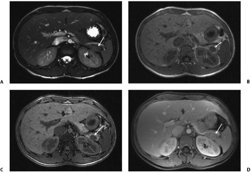
Fig. 41.1 (A) Axial T2-weighted image in a young man shows a well-defined hypointense lesion in the tail of the pancreas (arrow) with signal characteristics similar to the spleen. (B) Axial T1-weighted gradient echo in-phase image in the same patient shows a well-defined hypointense lesion in the tail of the pancreas (arrow), isointense to the spleen. (C) Axial T1-weighted gradient echo out-of-phase image in the same patient shows no drop of signal in the pancreatic tail lesion (arrow). (D) Axial postcontrast fat-saturated T1-weighted image in the same patient shows the homogeneously enhancing, isointense lesion in the tail of the pancreas (arrow), isointense to the spleen.
Radiologic Findings
Axial magnetic resonance (MR) images of the pancreas show the presence of a focal lesion in the tail of the pancreas that has signal characteristics similar to that of the spleen on T1- and T2-weighted images (Fig. 41.1).
Diagnosis
Accessory spleen in the pancreatic tail
Differential Diagnosis
- Pancreatic primary neoplasm
- Metastases
- Thrombosed aneurysm
- Hematoma
- Complex pseudocyst
- Abscess
Discussion
Background
Related posts:


Stay updated, free articles. Join our Telegram channel

Full access? Get Clinical Tree


