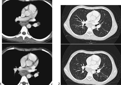
CASE 41 A 10-year-old recent immigrant presents with a 2-month history of intermittent fevers and cough. Figure 41A Frontal (Fig. 41A1) and lateral (Fig. 41A2) chest radiographs in a child with intermittent cough and fever reveals a small, right upper lobe peripheral focus of air-space density. Ipsilateral right superior hilar adenopathy is seen (see also Figs. 41B, 41C, and 41D). A linear opacity of thickened lymphatic channel is noted between the parenchymal focus and the regional adenopathy, constituting Ranke’s complex. Figure 41B Soft tissue (1) and lung windows (2) from a CT scan demonstrating similar findings as in Fig. 41A. There is mediastinal and hilar adenopathy, which demonstrate peripheral thick enhancement and central low attenuation, consistent with caseating necrosis. The lung windows demonstrate the small peripheral parenchymal focus and the thick line of lymphatic drainage leading to the hilum. Primary tuberculosis Differential diagnosis of primary tuberculous complex Tuberculosis is a disease undergoing an alarming, worldwide resurgence due to many factors, including the relatively recent HIV pandemic, the relative ease of worldwide travel, and the emergence of drug resistant forms. The World Health Organization estimated that 100,000 children worldwide died in 1998 from tuberculosis. Children constitute ~6% of the 22,000 new annual symptomatic cases in the United States.
Clinical Presentation
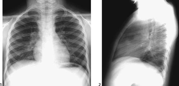
Radiologic Findings
Diagnosis
Differential Diagnosis
Discussion
Background
Etiology
Related posts:


Stay updated, free articles. Join our Telegram channel

Full access? Get Clinical Tree


