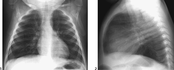
CASE 42 An 18-month-old girl presents with recurrent episodes of respiratory distress and suspected pneumonia. Figure 42A Frontal (Fig. 42A1) and lateral (Fig. 42A2) views of the chest in an 18-month-old girl reveal subtle early changes of lower airways disease, including mild diffuse overinflation and mild bilateral central bronchial wall thickening, consistent with early changes of cystic fibrosis. This was the third episode of suspected lower airway respiratory disease in the first year of life, prompting the child’s physician to perform a sweat test. Note that the area of subsegmental atelectasis is greater in the right upper lobe than in the left upper lobe. Early cystic fibrosis Cystic fibrosis is said to be the most common life-shortening inherited disease in the Caucasian population, affecting approximately 1 in 2500 live births in the United States and northern Europe. It is estimated that ~4% of whites are heterozygous for the most common form of the disorder. Current genetic technology has identified the genetic defect to be a mutation on the long arm of chromosome 7. The most common mutation is at the position dF 508 (70–80% of cases). The resultant mutation causes a physiologic disturbance in the sodium chloride ion transport system in epithelial cell membranes. This results in viscous secretions causing inspissated mucus in the tracheobronchial tree, digestive system, reproductive system, and sweat glands.
Clinical Presentation

Radiologic Findings
Diagnosis
Differential Diagnosis
Discussion
Background
Etiology
Clinical Findings
Stay updated, free articles. Join our Telegram channel

Full access? Get Clinical Tree


