Indication: Sanguineous secretion from the left nipple.
History: Breast conservation therapy of the right breast 7 years previously.
Risk profile: Increased by earlier occurrence of breast cancer.
Age: 67 years.
Fig. 43.1 Sonography.
Clinical Findings
No findings at palpation. Sanguineous secretion from one duct of the left breast.
Fig. 43.2a,b Digital mammography and galactography of the left breast, CC view.
Fig. 43.3a,b Digital mammography and galactography of the left breast, ML view.
Fig. 43.4 Magnification view of galactography of the left breast (CC).
Fig. 43.5a-c Contrast-enhanced MR mammography.
Fig. 43.6a-c Contrast-enhanced MR mammography.
Fig. 43.7 Contrast-enhanced MR mammography. Maximum intensity projection.
Fig. 43.8 Signal-to-time curves of the lesion in the left breast.
|
Please characterize ultrasound, mammography, and MRI findings. What is your preliminary diagnosis? What are your next steps? |
This is the imaging study of a symptomatic woman with a high risk profile presenting with pathological secretion from the left nipple.
Ultrasound
Following an ectatic duct in the lower inner quadrant of the left breast, ultrasound showed vague indications of a hypoechoicmass, 3 mm in diameter, with indeterminate distal signal behavior. US BI-RADS 3.
Mammography
Imaging showed inhomogeneous dense parenchyma, ACR type 3. There were no suspicious masses or densities, particularly in the lower inner quadrant of the left breast, and no microcalcifications. No architectural distortion. Galactography demonstrated a 3 mmcontrast-filling defect in the lower inner quadrant of the left breast. BI-RADS right 1/left 3. PGMI is not defined for unilateral imaging.
MR Mammography
Consistent with the lesion found in galactography, there was a hypervascularized, partially ill-defined lesion of 3 mm diameter in the lower inner quadrant of the left breast, with an increased signal in T2-weighted imaging. The lesion showed initial signal increase of 120% followed by washout.
MRI Artifact Category: 2
MRI Density Type: 1
MRM score |
Finding |
Points |
Shape |
round |
0 |
Border |
ill-defined |
1 |
CM Distribution |
homogenous |
0 |
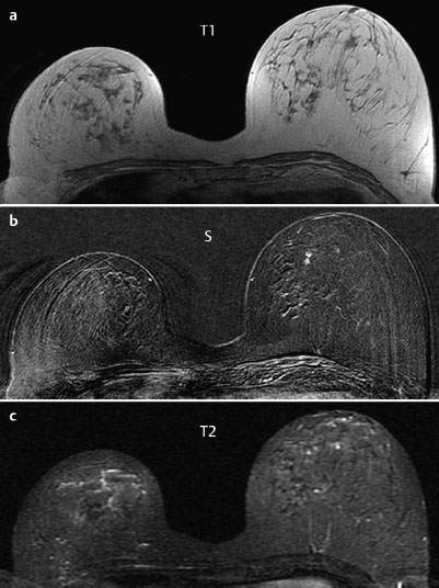
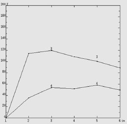
Initial Signal Intensity Increase |
strong |
2 |
Post-initial Signal Intensity Character |
wash-out |
2 |
MRI score (points) |
|
5 |
MRI BI-RADS |
4 |
Differential Diagnosis
Papilloma, adenoma, fibroadenoma, carcinoma.
BI-RADS Categorization | ||
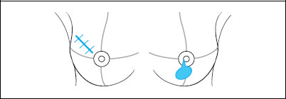
Clinical Findings |
right 1 |
left 3 |
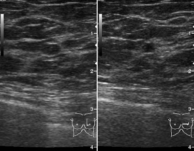
Ultrasound |
right 1 |
left 3 |
Mammography |
right 1 |
left 3 |
MR Mammography |
right 1 |
left 4 |
BI-RADS Total |
right 1 |
left 4 |
Procedure
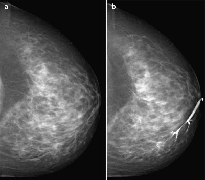
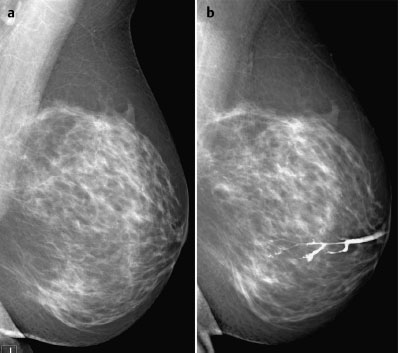
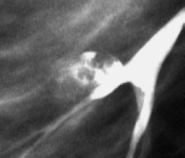
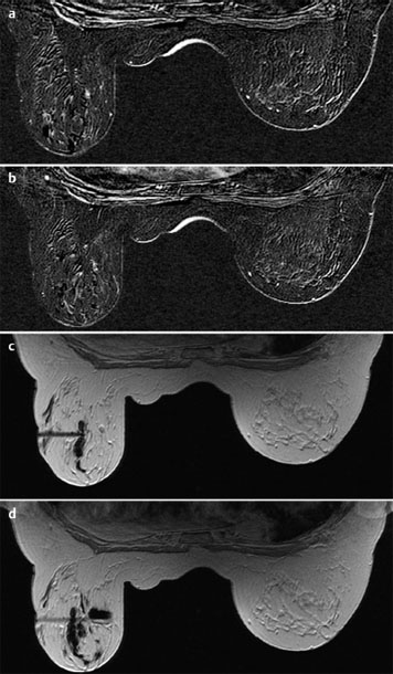
Histological analysis of the galactography findings in the lower inner quadrant of the left breast. MR-guided percutaneous vacuum biopsy with the objective of complete extraction of the lesion as a diagnostic and therapeutic intervention (Figs. 43.9 and 43.10).
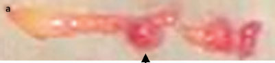
Fig. 43.10a First biopsy specimen containing a round nodular structure (arrow).
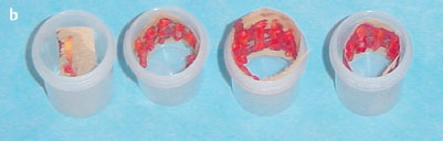
Fig. 43.10 b Complete set of 20 biopsy specimens ready for dispatch to histology lab.
Histology
Completely removed intraductal papilloma (extracted in the very first core specimen (see Fig. 43.10a). No malignancy.
Intraductal papilloma.

Stay updated, free articles. Join our Telegram channel

Full access? Get Clinical Tree

