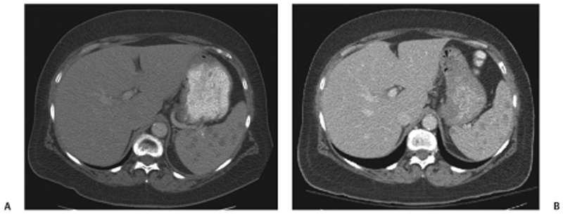
CASE 43 A 63-year-old man complains of fatigue, general malaise, and weight loss. Fig. 43.1 (A,B) Axial contrast-enhanced CT images of the abdomen show multiple small, hypodense nodular lesions scattered within a normal-sized spleen. No lesions are seen in the liver. There is no evidence of abnormal lymphadenopathy or fluid collection. Axial contrast-enhanced computed tomography (CT) images of the abdomen (Fig. 43.1) demonstrate multiple small, hypodense lesions within the splenic parenchyma. Splenic sarcoidosis
Clinical Presentation
Radiologic Findings
Diagnosis
Differential Diagnosis
Discussion
Background
Related posts:


Stay updated, free articles. Join our Telegram channel

Full access? Get Clinical Tree


