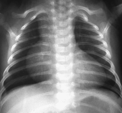
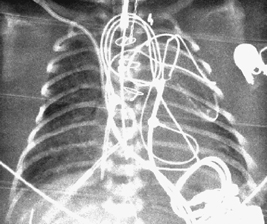
CASE 44 A newborn infant developed severe respiratory distress and cyanosis immediately after birth. Figure 44A A frontal chest radiograph (Fig. 44A) obtained immediately after birth demonstrates a markedly enlarged heart. The right atrial border extends to the upper mediastinum and bulges far to the right. The pulmonary vascularity is slightly prominent. Figure 44B Figure 44C Frontal chest radiograph of a newborn with pulmonary atresia and intact ventricular septum shows a markedly enlarged heart with an egg-on-side appearance. The right atrial contour is prominent, and the pulmonary vascularity is reduced. Figure 44D Frontal chest radiograph of a newborn with heart block shows severe cardiomegaly and severe pulmonary edema. Ebstein’s malformation of the tricuspid valve (Fig. 44B) with patent ductus arteriosus Severe cardiomegaly in the newborn:
Clinical Presentation
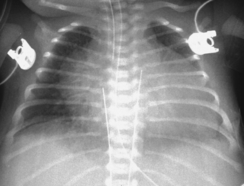
Radiologic Findings
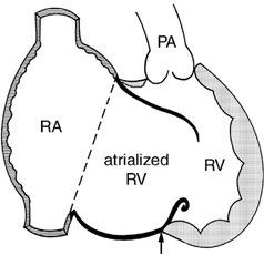
Diagnosis
Differential Diagnosis
Discussion
Clinical Findings
Related posts:


Stay updated, free articles. Join our Telegram channel

Full access? Get Clinical Tree


