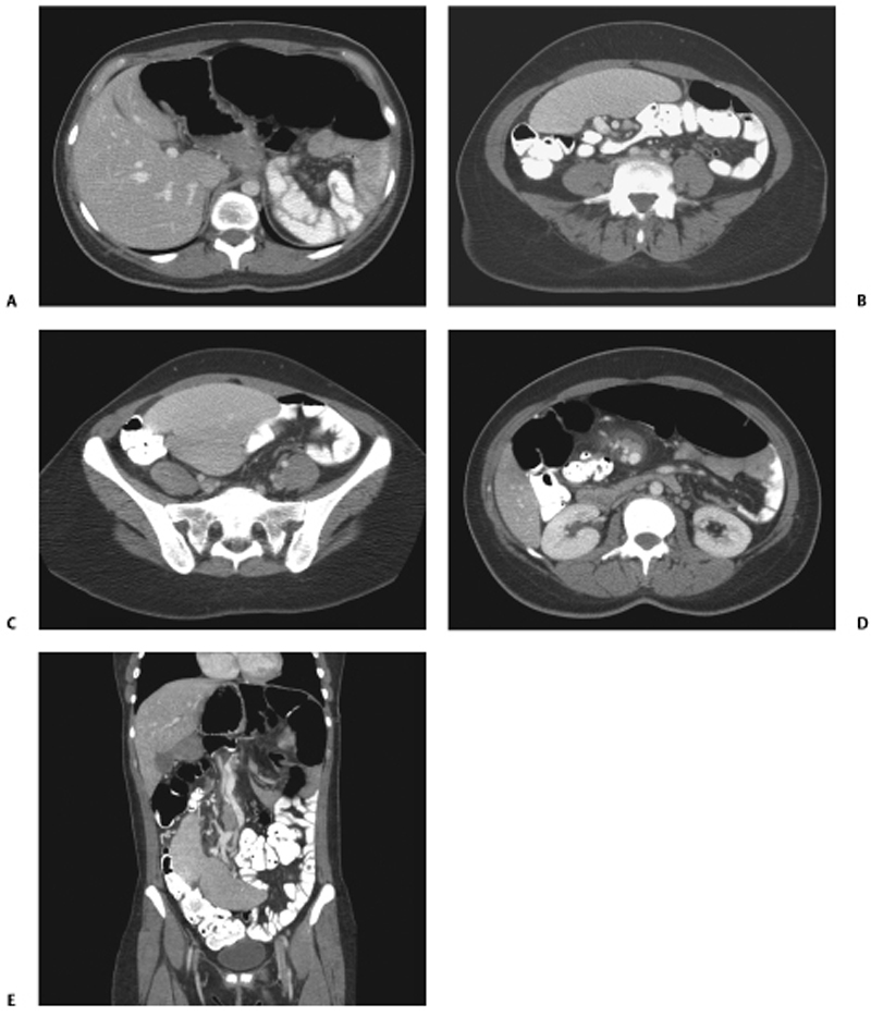
CASE 44 A 30-year-old woman complains of abdominal pain. Fig. 44.1 (A–E) Contrast-enhanced CT study shows the absence of the spleen in the left upper quadrant. The spleen is present inferior to the liver with a torsed pedicle seen as a whirl-like appearance of splenic vessels and surrounding fat noted at the splenic hilum (D). Axial contrast-enhanced computed tomography (CT) images of the abdomen (Fig. 44.1) reveal the absence of the spleen in the left upper quadrant. The spleen is seen inferior to the liver in the right lower quadrant. Wandering spleen This clinical entity usually presents between the ages of 20 and 40 years and is most commonly seen in women; most are of reproductive age at the time. Children make up one third of all cases, and 30% of them are younger than 10 years of age. This condition can be acquired or congenital. The acquired form is usually seen in multiparous women, attributed to hormonal changes during pregnancy that cause laxity of the ligaments normally attached to the spleen. In the congenital form, there is failure of normal development of the dorsal mesogastrium when the lesser sac is formed. The attachments of the dorsal mesentery to the posterior peritoneum and diaphragm are faulty. Suspensory ligaments of the spleen are not formed or are only partially formed. The length of its vascular pedicle determines the mobility of the spleen in the absence of some or all of these ligaments.
Clinical Presentation

Radiologic Findings
Diagnosis
Differential Diagnosis
Discussion
Background
Clinical Findings
Stay updated, free articles. Join our Telegram channel

Full access? Get Clinical Tree


