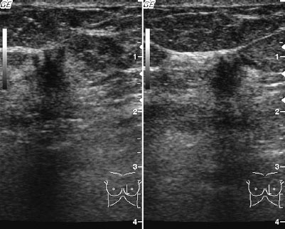
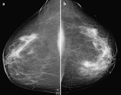
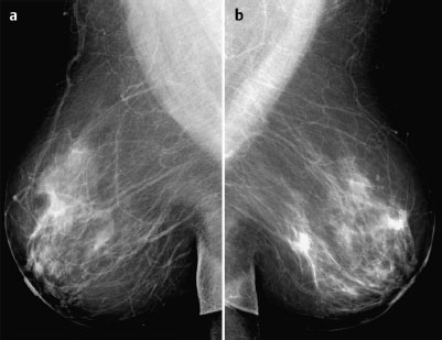
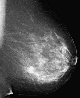
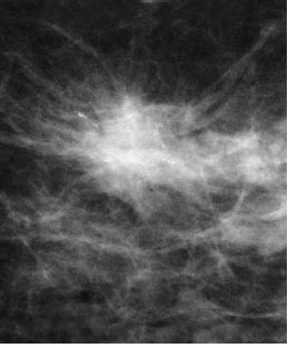
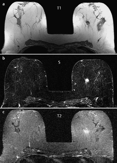
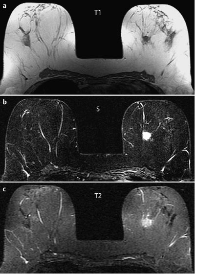
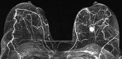
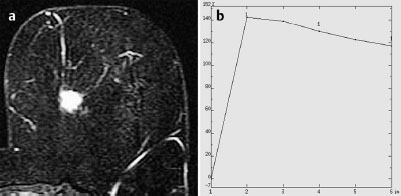
Case 45 Indication: Screening. History: Unremarkable. Risk profile: No increased risk. Age: 58 years. Fig. 45.1 Ultrasound. No suspect findings. Fig. 45.2a,b Digital mammography, CC view. Fig. 45.3a,b Digital mammography, MLO view. Fig. 45.4 Digital mammography, left breast. ML view. Fig. 45.5 Magnification view, left breast (CC). Fig. 45.6a-c Contrast-enhanced MRI of the breasts. Fig. 45.7a-c Contrast-enhanced MRI of the breasts. Fig. 45.9a,b Signal-to-time curve. Please characterize ultrasound, mammography, and MRI findings. What is your preliminary diagnosis? What are your next steps? This case demonstrates the imaging studies of an asymptomatic woman. Ultrasound showed an irregular hypoechoic lesion (diameter1 cm) with distal shadowing and architectural distortion in the lower inner quadrant of the left breast. US BI-RADS 5. The parenchyma was bilaterally symmetric with fibroglandular texture, ACR type 2. A hyperdense, lobulated mass of 1 cm diameter with spiculation and predominantly monomorphous microcalcifications was visible in the left lower inner quadrant. Mammograms showed no architectural distortion (BI-RADS right 1/left 5). PGMI: CC view P; MLO view G (inframammary fold). MRI demonstrated a partially lobulated, partially spiculated, homogeneously enhancing mass in the lower inner quadrant of the left breast with a diameter of 1 cm. This mass had an initial signal increase of 115% and postinitial washout as well as increased signal in T2-weighted imaging. MRI Artifact Category: 1 MRI Density Type: 1
Clinical Findings

Ultrasound
Mammography
MR Mammography
MRM score | Finding | Points |
Shape | round | 0 |
Border | spiculated | 1 |
CM Distribution | inhomogenous | 1 |
Initial Signal Intensity Increase | strong | 2 |
Post-initial Signal Intensity Character | wash-out | 2 |
MRI score (points) |
| 6 |
MRI BI-RADS |
| 5 |
 Preliminary Diagnosis
Carcinoma.
Clinical Findings | right 1 | left 1 |
Ultrasound | right 1 | left 5 |
Mammography | right 1 | left 5 |
MR Mammography | right 1 | left 5 |
BI-RADS Total | right 1 | left 5 |
Procedure
Ultrasound guided core biopsy of the lesion in the left breast for histological analysis.
Histology
Lobular invasive carcinoma.
Further procedure
Resection of tumor after preoperative hook-wire localization (Fig. 45.10a, b). Perioperative specimen radiography to verify complete excision of tumor (Fig. 45.11).
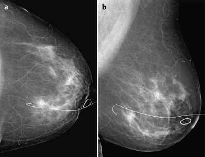
Fig. 45.11 Specimen radiography.
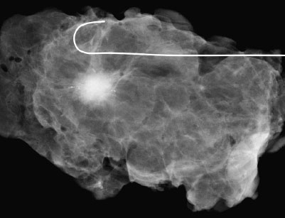
Fig. 45.10a,b Hook-wire localization seen in mammography.
Histology
Lobular invasive breast carcinoma with a diameter of 12 mm. Axillary lymph node status normal.
ILC pTIc, pN0, G2.
Stay updated, free articles. Join our Telegram channel

Full access? Get Clinical Tree


