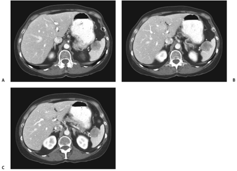
CASE 45 A 61-year-old woman with a previous history of endometrial carcinoma and elastofibroma of the shoulder complains of upper abdominal pain. She underwent splenectomy following her initial evaluation. Fig. 45.1 (A–C) Axial contrast-enhanced CT images of the abdomen show an enhancing, exophytic lesion adjacent to the splenic hilum. Axial contrast-enhanced computed tomography (CT) images of the abdomen (Fig. 45.1) show a heterogeneously enhancing, exophytic lesion adjacent to the splenic hilum. Inflammatory splenic pseudotumor
Clinical Presentation

Radiologic Findings
Diagnosis
Differential Diagnosis
Discussion
Background
Stay updated, free articles. Join our Telegram channel

Full access? Get Clinical Tree


