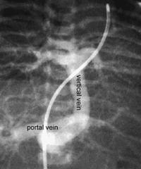
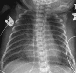
CASE 45 A term newborn developed severe hypoxemia, cyanosis, and tachypnea immediately after birth. The obstetric history was unremarkable. Figure 45A A frontal chest radiograph (Fig. 45A) shows moderately hyperinflated lungs with generalized increase in interstitial lung markings. The heart is normal in size and configuration. Figure 45B Injection into the main pulmonary artery at cardiac catheterization shows that all pulmonary veins make a confluence at a vertical vein that connects to the portal vein in the liver. Figure 45C Plain chest radiograph, of a patient with hypoplastic left heart syndrome and restrictive atrial communication, shows generalized increase in interstitial lung markings and air trapping as in Fig. 45A. Total anomalous pulmonary venous connection (TAVPC) to the portal vein (Fig. 45B)
Clinical Presentation
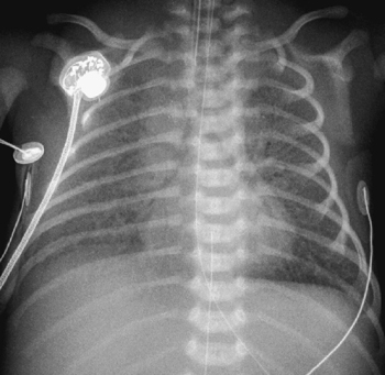
Radiologic Findings
Diagnosis
Differential Diagnosis
Discussion
Clinical Findings
Related posts:


Stay updated, free articles. Join our Telegram channel

Full access? Get Clinical Tree


