Case 46
 Clinical Presentation
A 72-year-old man with hematuria.
 Imaging Findings
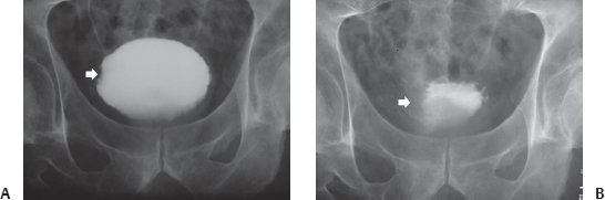
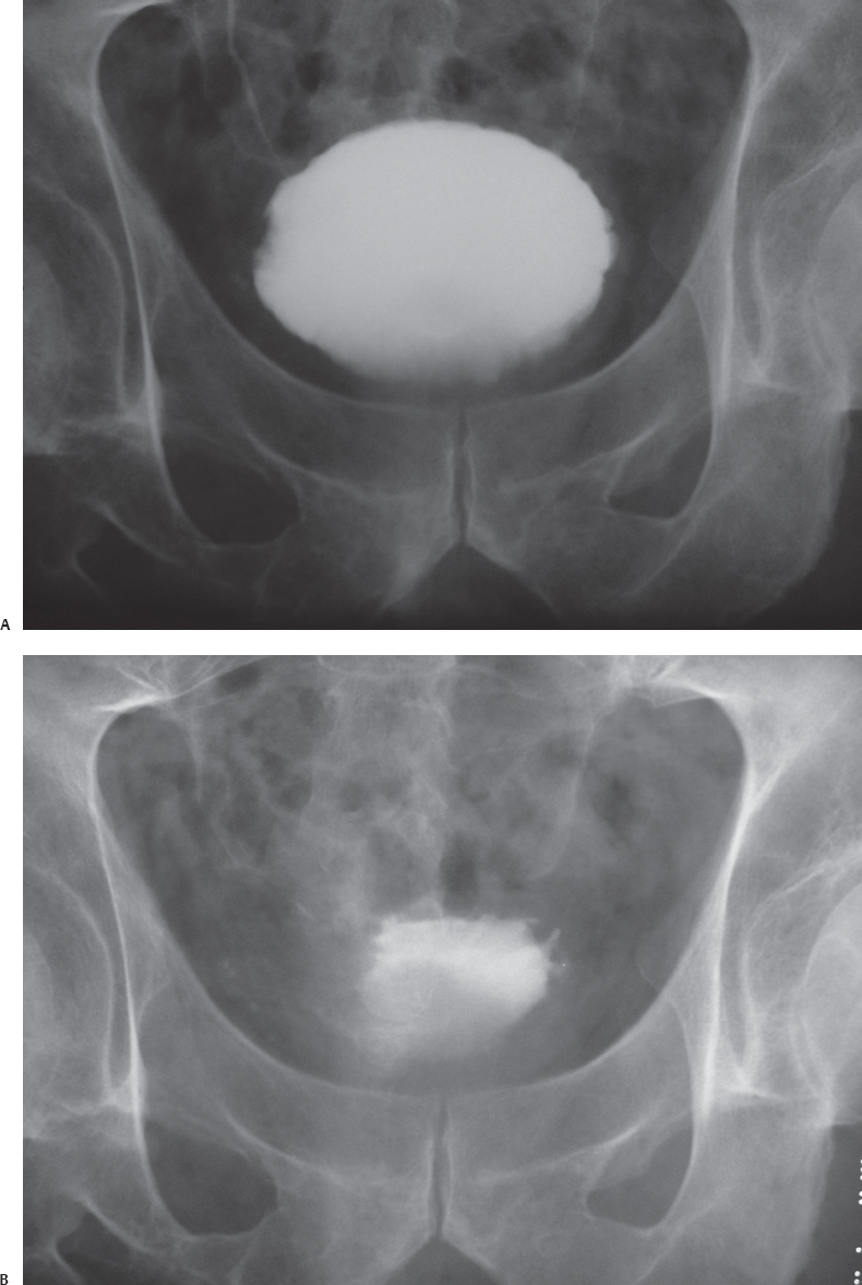
(A) Full-bladder image from an intravenous pyelogram (IVP) shows a subtle filling defect (arrow) on the right wall of the urinary bladder. The bladder base appears to be lifted by a prostatic impression. (B) Postvoid bladder image from the same IVP study shows that the filling defect (arrow) on the right wall of the urinary bladder is much more prominent, creating asymmetry of the urinary bladder, which has an irregular outline. The bladder mucosa is normal. No significant residual urine is seen.
 Differential Diagnosis
• Urinary bladder cancer: This is characterized by an irregular filling defect of the bladder wall that is partially obscured by contrast in the full bladder.
• Blood clot in the urinary bladder: This is also more conspicuous in a postvoid image. However, it is often mobile and smooth.
• Extrinsic impression on the urinary bladder: This is more conspicuous when the bladder is full.
Stay updated, free articles. Join our Telegram channel

Full access? Get Clinical Tree


