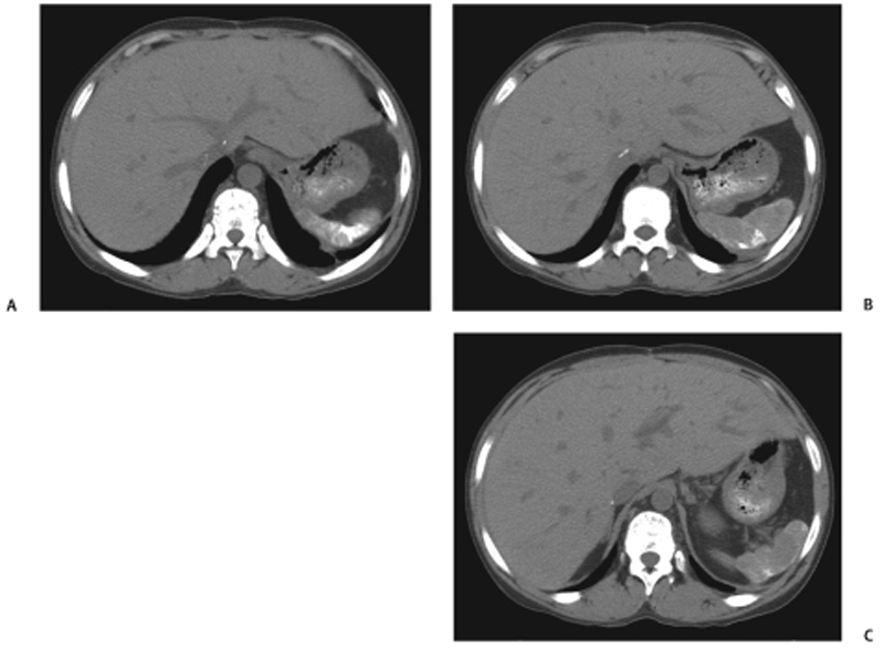
CASE 46 A 25-year-old patient presents with sickle cell disease. Fig. 46.1 (A–C) Noncontrast CT scans of the upper abdomen show a partially calcified small spleen. Noncontrast axial computed tomography (CT) scan shows a calcified small spleen (Fig. 46.1). Autosplenectomy from sickle cell disease
Clinical Presentation
Radiologic Findings
Diagnosis
Differential Diagnosis
Discussion
Background
Related posts:


Stay updated, free articles. Join our Telegram channel

Full access? Get Clinical Tree


