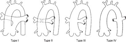
CASE 46 A newborn presents with mild cyanosis and failure to thrive. Figure 46A The frontal chest radiograph (Fig. 46A1) shows a moderately enlarged heart with an egg-on-side configuration and increased pulmonary vascularity. The superior mediastinum is narrow because of thymic hypoplasia. Note that the anterior upper mediastinum is empty in lateral view (Fig. 46A2). Figure 46B Schematic diagram of four types of truncus arteriosus as described by Collet and Edwards. Truncus arteriosus with DiGeorge syndrome Most patients with truncus are recognized as having congenital heart disease during the neonatal period. During the first few weeks of life, persistence of increased pulmonary vascular resistance results in mild cyanosis with little evidence of heart failure. As the pulmonary vascular resistance decreases, cyanosis may disappear and be replaced by signs of congestive heart failure. When the truncal valve is severely regurgitant, the signs of congestive heart failure may manifest immediately after birth. Generally, the heart is overactive, and a pansystolic murmur is heard at the left precordium. Microdeletion of chromosome 22q11 is seen in ~40% of patients with truncus, and patients with this deletion include DiGeorge and velocardiofacial syndrome cases.
Clinical Presentation
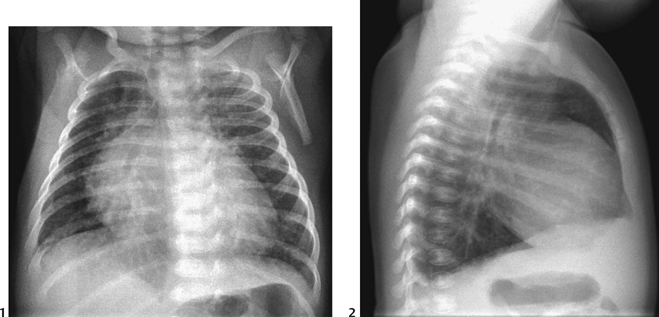
Radiologic Findings
Diagnosis
Differential Diagnosis
Discussion
Clinical Findings
Pathology
Related posts:


Stay updated, free articles. Join our Telegram channel

Full access? Get Clinical Tree


