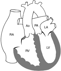
CASE 47 A newborn presents with cyanosis and tachypnea. Figure 47A A frontal chest radiograph (Fig. 47A) shows a mildly enlarged heart with an egg-on-side configuration and mildly increased interstitial markings. Figure 47B T1-weighted coronal MRI (1) of a patient with complete transposition of the great arteries shows an egg-on-side appearance, as shown in 2. Ao, aorta; LV, left ventricle; RV, right ventricle. Figure 47C Diagram showing complete transposition of the great arteries. Ao, aorta; LA, left atrium; LV, left ventricle; PA, pulmonary artery; RA, right atrium; RV, right ventricle. Complete transposition of the great arteries. A T1-weighted MRI in coronal plane (Fig. 47B1) shows that the aorta arises from the morphologically right ventricle. Note that the cardiac configuration in both plain radiograph and MRI is egg-shaped (Fig. 47B2). Egg-on-side appearance: The circulatory arrangement of complete transposition is such that all patients have significant desaturation from birth. Cyanosis is usually evident from the first day of life when the ventricular septum is intact or within a week or month when there is a large ventricular septal defect. Transposition of the great arteries is defined as discordant connection between the ventricles and great arteries; that is, the morphologic right ventricle connects to the aorta and the left ventricle to the pulmonary artery. Complete transposition of the great arteries is defined as the combination of normal concordant atrioventricular connection and discordant ventriculoarterial connection (Fig. 47C
Clinical Presentation
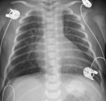
Radiologic Findings

Diagnosis
Differential Diagnosis
Discussion
Clinical Findings
Pathology
![]()
Stay updated, free articles. Join our Telegram channel


Full access? Get Clinical Tree


