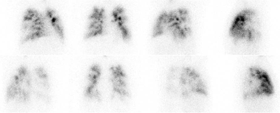
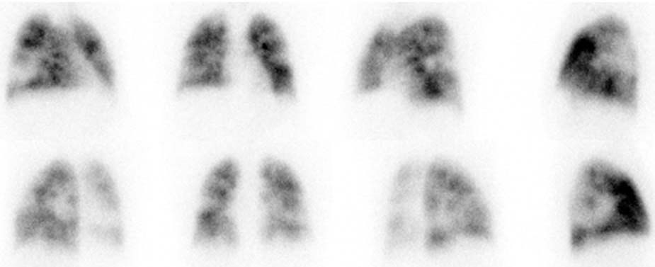
CASE 47 A 50-year-old man with a productive cough presents with atypical chest pain. Cardiac catheterization is negative, and a chest radiograph shows bilateral patchy opacities in both lungs. Fig. 47.1 Fig. 47.2 • A 40.0 mCi dose of 99mTc-DTPA is placed in the aerosolizer (delivers < 1 mCi to the lungs). • Use a low-energy, all-purpose collimator. • Energy window 20% centered at 80 keV. • Views are anterior, right anterior oblique, left anterior oblique, posterior, right posterior oblique, left posterior oblique, right lateral view, and left lateral. • A 3.0 mCi dose of 99mTc-MAA is administered intravenously with the patient supine. • The patient should cough and take several deep breaths before administration of the MAA to clear any areas of resting atelectasis. • The patient should breathe normally during tracer injection. • Use a low-energy, all-purpose collimator. • Energy window 20% centered at 140 keV. • Imaging time is 500,000 counts per view. • Matrix size is 128 × 128. • Views are anterior, right anterior oblique, left anterior oblique, posterior, right posterior oblique, left posterior oblique, right lateral view, and left lateral. The lung ventilation images demonstrate large defects that do not appear to correspond to the segmental anatomy of the lung (Fig. 47.1
Clinical Presentation
Technique
Lung Ventilation Scan
Lung Perfusion Scan
Image Interpretation
![]()
Stay updated, free articles. Join our Telegram channel

Full access? Get Clinical Tree


