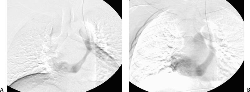
CASE 47 A 35-year-old asymptomatic patient presented to interventional radiology for placement of a permanent catheter for hemodialysis. Because the patient had exhausted options for arteriovenous shunt placement in the left arm, the left internal jugular vein was chosen over the right for catheterization. Figure 47-1 Persistent left superior vena cava. (A) Venography performed from the left internal jugular vein shows absence of the left brachiocephalic vein and anomalous drainage into the coronary sinus. (B) Eventual drainage into the right atrium and pulmonary arteries. After catheterization and wire advancement into the left internal jugular vein, the radiologist was unable to access the left brachiocephalic vein. Venography performed in the frontal projection showed central venous drainage to the right atrium via an anomalous vessel projected over the left mediastinum (Fig. 47-1). Persistent left superior vena cava (PLSVC) draining into the coronary sinus. The patient underwent placement of a permanent hemodialysis catheter via the right internal jugular vein. Figure 47-2 A 62-year-old male after left and right central line placement in the intensive care unit. Portable chest radiograph in the frontal projection shows a left mediastinal catheter inadvertently placed into the aorta via the carotid artery.
Clinical Presentation
Radiologic Studies
Diagnosis
Treatment

Discussion
Background
Related posts:


Stay updated, free articles. Join our Telegram channel

Full access? Get Clinical Tree


