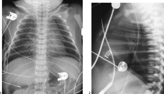
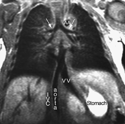
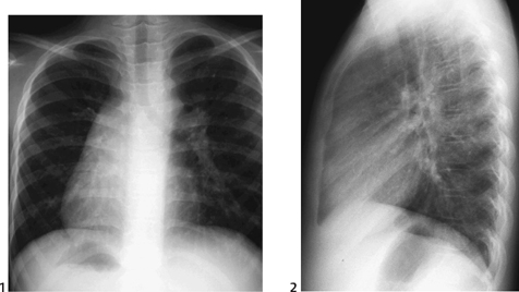
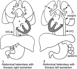
CASE 48 A newborn presents with cyanosis and tachypnea. Figure 48A A frontal chest radiograph (Fig. 48A1) shows the stomach bubble in a left paramedian location and the transverse liver. There is a levocardia. In the lateral chest radiograph (Fig. 48A2), the upper lobar bronchi are located in similar horizontal levels. The pulmonary arterial shadow is seen mostly in front of the airway. Both lungs show increased interstitial markings due to pulmonary venous hypertension secondary to obstructive type of total anomalous pulmonary venous connection. Figure 48B T1-weighted coronal MRI shows right-isomeric branching pattern of the bronchi (arrows), juxtaposition of the aorta and inferior vena cava (IVC) on the right side, and a left-sided stomach. All pulmonary veins make a confluence at a vertical vein (VV) that drains into the portal vein. AA, aortic arch. Figure 48C Chest radiographs in a patient with left isomerism. The frontal view (1) shows symmetrically long bronchi, right-sided stomach, and levocardia. The lateral view (2) shows end-on shadows of both upper lobar bronchi in similar horizontal levels. The pulmonary arteries are seen above and behind the upper lobar bronchi. Right isomerism with transverse liver and asplenia. A T1-weighted MRI (Fig. 48B) shows symmetrically short bronchi. Neither right nor left pulmonary artery is above the main bronchus. The abdominal aorta and inferior vena cava are juxtaposed on the right side of the spine. The pulmonary veins make a confluent channel to connect to the portal vein via a vertical vein. Figure 48D Diagrams show visceral heterotaxy with thoracic right isomerism and asplenia (1) and visceral heterotaxy with thoracic left isomerism and polysplenia (2) gastric bubble; IVC, inferior vena cava; PA, pulmonary artery; SVC, superior vena cava
Clinical Presentation
Radiologic Findings
Diagnosis
Differential Diagnosis
Discussion
Clinical Findings
Related posts:


Stay updated, free articles. Join our Telegram channel

Full access? Get Clinical Tree


