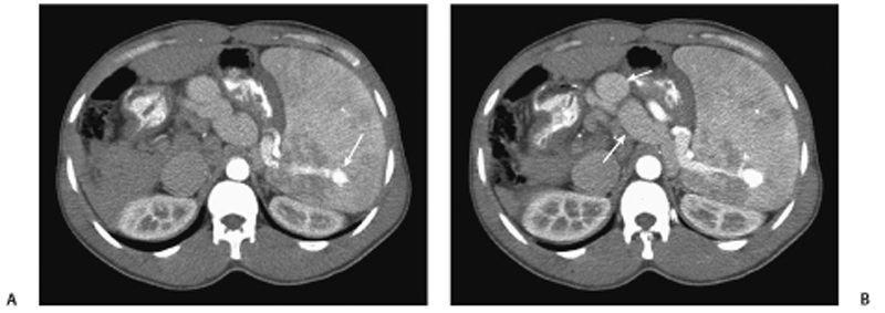
CASE 48 A 40-year-old man presents with abdominal pain and a single episode of an upper gastrointestinal (GI) bleed. Fig. 48.1 (A) Contrast-enhanced axial CT image shows the presence of a splenic arteriovenous fistula (arrow). (B) Note the dilated upper abdominal venous varices (arrows). Contrast-enhanced axial computed tomography (CT) scans (Fig. 48.1) show an enlarged spleen with an arteriovenous (AV) fistula and early opacification of the splenic vein. Note the enlarged collateral veins in the upper abdomen. Splenic AV fistula with features of portal hypertension
Clinical Presentation
Radiologic Findings
Diagnosis
Differential Diagnosis
Discussion
Background
Related posts:


Stay updated, free articles. Join our Telegram channel

Full access? Get Clinical Tree


