CASE 49 A 15-year-old girl presents with exertional dyspnea and exercise intolerance. Figure 49A A frontal chest radiograph (Fig. 49A1) at presentation shows mild cardiomegaly, convex bulging of pulmonary arterial segment of the left heart border. The central pulmonary arteries are dilated, whereas peripheral pulmonary vessels are attenuated. The lateral chest radiograph (Fig. 49A2) also shows central-peripheral discrepancy in pulmonary artery size. The retrosternal space is obliterated because of right ventricular hypertrophy. Primary pulmonary hypertension The clinical features of primary pulmonary hypertension vary with the severity of the lesion. Patients frequently complain of unusual fatigue, shortness of breath, and chest pain on minor exertion. The chest pain is attributable to increased myocardial oxygen demand from increased right ventricular workload. Some patients present with episodes of fainting or loss of consciousness. On physical examination, the most consistent finding is an increased pulmonic component of the second heart sound. Elevation of jugular venous pulsation and ankle swelling are secondary findings of right heart failure.
Clinical Presentation
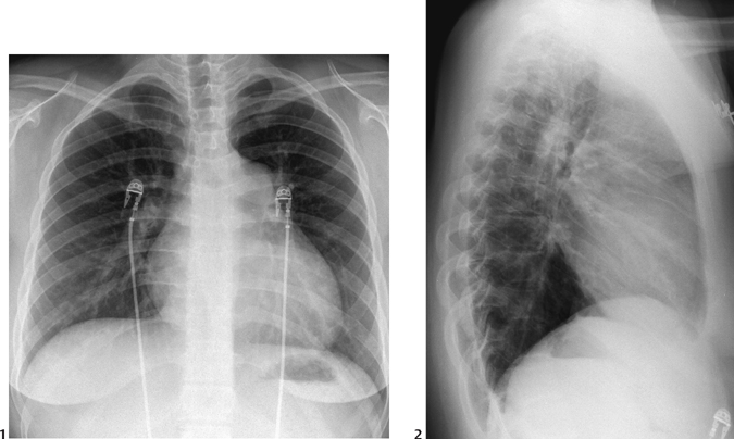
Radiologic Findings
Diagnosis
Differential Diagnosis
Discussion
Clinical Findings
Pathophysiology
Related posts:


Stay updated, free articles. Join our Telegram channel

Full access? Get Clinical Tree


