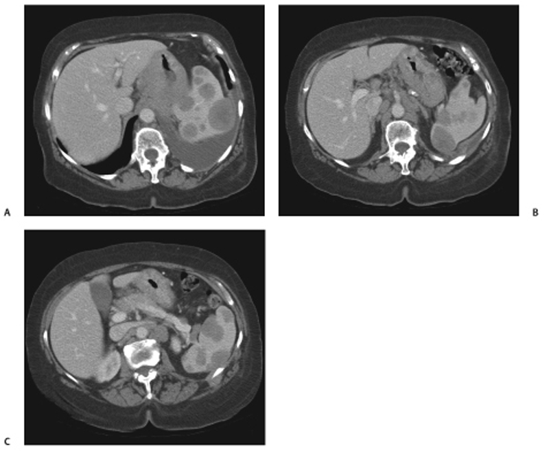
CASE 49 A 55-year-old woman presents with abdominal pain, low-grade fever, and weight loss. Fig. 49.1 (A–C) Axial contrast-enhanced CT images show multiple, well-defined, low-density focal lesions in the spleen. Also seen are thickening of the stomach wall, enlarged retroperitoneal abdominal lymph nodes, and a left pleural effusion. Contrast-enhanced axial computed tomography (CT) scans show multiple low-density lesions in the spleen, along with diffuse thickening of the stomach, left pleural effusion, and enlarged abdominal lymph nodes (Fig. 49.1). Secondary involvement of the spleen in lymphoma
Clinical Presentation
Radiologic Findings
Diagnosis
Differential Diagnosis
Discussion
Related posts:


Stay updated, free articles. Join our Telegram channel

Full access? Get Clinical Tree


