5 Skull Base and Temporal Bone
The anterior skull base consists of the cribriform plate of the ethmoid bone centrally, the orbital plates of the frontal bone laterally, the lesser wings of the sphenoid bone, and the planum sphenoidale (presphenoid) posteriorly. The superior surface of the anterior skull base forms the floor of the anterior cranial fossa; the inferior surface constitutes the roof of the nasal cavity, frontal and ethmoid sinuses (fovea ethmoidalis), and orbits. In addition to the frontal, ethmoid, and sphenoid bones, the undersurface of the anterior skull base is formed by the maxilla, vomer, palatine, zygomatic bones, and paired pterygoid processes, extending inferiorly from the sphenoid body. The cribriform plate contains multiple perforations through which the sensory nerve fibers of the olfactory nerve (cranial nerve [CN] I) and ethmoid arteries pass. The anterior ethmoidal foramen is located just anterior to the cribriform foramina and transmits the anterior ethmoidal artery, vein, and nerve. The posterior ethmoidal foramen is located just posterior to the cribriform foramina and transmits the posterior ethmoidal artery, vein, and nerve. The foramen cecum, located in the mid-line, anterior to the crista galli, transmits small vessels and is occasionally the starting point of cephaloceles or nasal gliomas.
The central skull base is formed by the sphenoid bone (basisphenoid and greater wings of the sphenoid) and the paired temporal bones. The middle cranial fossa extends from the lesser wings of the sphenoid and the frontal bones to the dorsum sellae and posterior clinoid processes medially and the superior margin of the petrous ridges laterally. Superior relationships of the middle cranial fossa are the temporal lobes, pituitary gland, cavernous sinus, Meckel cave, and CN II to VI. The floor of the sphenoid sinus and the basisphenoid form the anterior roof of the pharyngeal mucosal space. The deep facial spaces that abut the exocranial surface of the central skull base are the retropharyngeal space and the perivertebral space and the paired parapharyngeal space, masticator space, carotid space, and parotid space.
The optic canal is a round aperture within the lesser wing of the sphenoid at its junction with the sphenoid body, which transmits the optic nerve and ophthalmic artery, both of which are contained in a dural sheath. Inferolaterally, the canal is separated from the superior orbital fissure by the inferior root of the lesser wing (“optic strut”).
The superior orbital fissure, formed by the cleft between the greater sphenoid wing inferolaterally and the lesser sphenoid wing superolaterally and the sphenoid body medially, transmits CN III, IV, V1, and VI, recurrent branches of the lacrimal artery, the orbital branch of the middle meningeal artery, and the superior ophthalmic vein.
The inferior orbital fissure, which transmits the infraorbital artery, vein, and nerve, is formed by the cleft between the body of the maxilla and the greater wing of the sphenoid bone. The inferior orbital fissure communicates inferiorly with the pterygopalatine fossa.
The foramen rotundum, actually a canal in the base of the greater sphenoid wing, just inferiorly and laterally to the superior orbital fissure and superolaterally to the vidian canal, transmits CN V2, the artery of the foramen rotundum, and emissary veins. The foramen rotundum empties anteriorly into the pterygopalatine fossa, which connects laterally with the masticator space through the pterygomaxillary fissure. Malignant tumors of the skin of the cheek, orbit, and sinonasal area may all use CN V2 as a perineural route to gain intracranial access.
The foramen ovale, which lies completely within the greater wing of the sphenoid, transmits CN V3 (from the middle cranial fossa to the masticator space), the lesser petrosal nerve, the accessory meningeal branch of the maxillary artery, and the emissary vein. Endocranially, the foramen ovale lies anteromedial to the foramen spinosum and posterolateral to the foramen rotundum. Exocranially, the foramen ovale is located at the base of the lateral pterygoid plate. Perineural tumor extension on the mandibular division of the trigeminal nerve in the masticator space may traverse the skull base through the foramen ovale, spread intracranially through Meckel cave and the preganglionic segment of the trigeminal nerve, and finally reach the pons at the root entry zone.
The foramen spinosum, located at the posteromedial aspect of the greater sphenoid wing, endocranially posterolateral to the foramen ovale, and exocranially anterior and lateral to the eustachian tube, transmits the middle meningeal artery and vein, as well as the meningeal branch of CN V3.
The vidian canal, situated in the body of the pterygoid plates below and inferomedial to the foramen rotundum in the body of the sphenoid bone, connects the pterygopalatine fossa anteriorly to the foramen lacerum posteriorly and transmits the vidian artery and nerve.
The foramen lacerum, located at the base of the medial pterygoid plate, bound anterolaterally by the greater wing of the sphenoid bone, posteriorly by the petrous apex, and medially by the sphenoid body and basiocciput, is not a true foramen but is a canal largely filled with fibrocartilage. It represents the cartilaginous floor of the anteromedial horizontal segment of the petrous internal carotid artery canal. An inconstant meningeal branch of the ascending pharyngeal artery and the vidian nerve may pierce the cartilage.
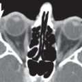
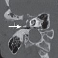
The sphenopalatine foramen, located in the high posterolateral wall of the nose, connects the lateral nasal cavity with the pterygopalatine fossa. The foramen transmits the lateral nasal and nasopalatine nerves and vessels. Nasal infection and tumors can access the intracranial space, orbit, and masticator space through this escape hatch.
The posterior skull base is made up of the sphenoid bone, temporal bones posterior to the petrous ridge, and occipital bones. The anterior portion of the posterior cranial fossa is formed by the clivus, which is derived from the fusion of the basisphenoid and the basiocciput. It extends from the dorsum sellae to the foramen magnum. The lateral wall of the posterior cranial fossa is formed superiorly by the posterior surface of the petrous temporal bone and inferiorly by the condylar part of the occipital bone. The posterior portion of the posterior cranial fossa is made up of the mastoid portion of the temporal bone and the squamous portion of the occipital bone. The superior surface of the posterior skull base forms the floor of the posterior cranial fossa; the inferior surface constitutes the posterior roof of the pharyngeal mucosal space; the carotid, parotid, retropharyngeal, and perivertebral spaces; and the cervical spine.
The foramen magnum is bound by the four segments of the occipital bone: the basiocciput anteriorly, the two parts of the exoocciput laterally, and the supraocciput posteriorly. The bones surrounding the foramen magnum serve as a site of attachment for numerous ligaments (apical, dental, and alar ligaments, the upper band of the cruciform ligament, the posterior longitudinal ligament, and the tectorial membrane) that stabilize the craniocervical junction. Through the foramen magnum pass the medulla oblongata, the meninges, the vertebral arteries, the anterior and posterior spinal arteries, the spinal accessory nerve (CN XI), and the veins that communicate with the internal vertebral venous plexus.
The hypoglossal canal traverses the occipital condyle ante-rolaterally while transmitting the hypoglossal nerve (CN XII), a meningeal branch of the ascending pharyngeal artery, and a venous plexus. The canal also transmits the rare persistent hypoglossal artery when it is present.
The common skull base apertures and their contents are summarized in Table 5.1. Diseases of the skull base can be intrinsic to the area or affect the skull base from either above or below. All these lesions are covered in other sections of this text: intracranial lesions that involve the skull base from above (“top-down” lesions) are discussed in Section I (Brain), lesions originating in the skull base in Section IV (Musculoskeletal System), and extracranial lesions affecting the skull base from below (“bottom-up” lesions) in Chapter 7 (Nasal Cavity and Paranasal Sinuses) and Chapter 8 (Suprahyoid Neck) of this section. All major lesions involving the skull base, with emphasis on intrinsic lesions, are summarized in Table 5.2.
Related posts:
Stay updated, free articles. Join our Telegram channel

Full access? Get Clinical Tree


