9 Infrahyoid Neck(Table 9.5 – Table 9.8)
Disease | CT Findings | Comments |
Pseudomass | ||
Levator claviculae muscle | This muscle has its origin in the upper cervical spine and inserts into the middle or lateral third of the clavicle and appears as nodular soft tissue structure in the posterior cervical space. | Normal anatomical variant, occurring in 2% to 3% of the population. Rarely, the muscle itself causes the impression of a posterior neck mass lesion. |
Congenital/developmental lesions | ||
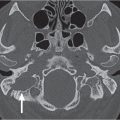
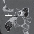
Third branchial cleft cyst | Rounded or ovoid, sharply marginated lesion in the posterior cervical space with central fluid density. The cyst wall is nearly imperceptible. The sternocleidomastoid muscle is usually laterally displaced. May contain air if the cyst communicates with the pyriform sinus via patent tract. When infected, the cyst wall may become thickened and irregular, the cyst content develops higher attenuation, and the surrounding fat planes may become obscured. | Rare lesion (accounts for only 3% of all branchial anomalies). May occur anywhere along the course of the third branchial cleft or pouch. Presents in adulthood as painless fluctuant mass in the posterior triangle of the neck. May be a ssociated with third branchial cleft sinus or fistula. |
Venous vascular malformation (cavernous hemangioma) | Lobulated soft tissue mass, isodense to muscle, containing rounded calcifications (phleboliths). Contrast enhancement may be patchy and delayed or homogeneous and intense. Fat hypertrophy in adjacent soft tissues may be present. | Vascular malformations are not tumors but true congenital low-flow vascular anomalies, have an equal gender incidence, may not become clinically apparent until late infancy or childhood, virtually always grow in size with the patient during childhood, and do not involute spontaneously. Vascular malformations are further subdivided into capillary, venous, arterial, lymphatic, and combined malformations. Venous vascular malformations, usually present in children and young adults, are the most common vascular malformations of the head and neck. The buccal region and dorsal neck are the most common locations. Frequently, venous malformations do not respect fascial boundaries and involve more than one deep fascial space (transspatial disease). They are soft and compressible. Lesion may increase in size with Valsalva maneuver, bending over, or crying. Swelling and enlargement can occur following trauma or hormonal changes (e.g., pregnancy). Pain is common and often caused by thrombosis. |
Lymphatic malformation (cystic hygroma, lymphangioma) | Thin-walled, multiseptated or unilocular, nonenhancing cystic mass that may insinuate in and around normal structures. Fluid–fluid levels may be present due to hemorrhage. | Lymphatic malformations represent a spectrum of congenital low-flow vascular malformations, differentiated by size of dilated lymphatic channels. Macrocystic lymphatic malformation is the most common subtype. Ninety percent of lymphatic malformations become clinically apparent by 3 y of age, whereas the remaining 10% present as neck mass in the young adult. May be sporadic or part of congenital syndromes (Turner, Noonan, and fetal alcohol syndromes). Most lymphatic malformations arise in the posterior cervical space and supraclavicular region. Up to 10% of all cervical cystic hygromas extend into the mediastinum. Rapid enlargement of a lymphangioma is usually due to hemorrhage into the cystic spaces of the mass. |
Inflammatory/infectious conditions | ||
Reactive lymphadenopathy | In reactive adenopathy, cervical lymph nodes may by definition enlarge to a maximum diameter of 1.5 cm but maintain their normal oval shape, isodense to muscle, and their homogeneous internal architecture with variable, usually mild enhancement. Enhancing linear markings within the node may be seen. Multiple nodal locations are typically present. Postinflammatory fatty infiltration appears as low-density nodal hilus (which is peripheral rather than central), mimicking necrosis in a node with pronounced lima bean shape (fat usually has a lower attenuation than tumor necrosis). | Reactive hyperplasia is a nonspecific lymph node reaction, current or prior to any inflammation in its draining area. Can occur at any age but is most common in the pediatric age group. |
Suppurative lymphadenitis | Enlarged node with internal low attenuation (intra-nodal abscess) and ringlike contrast enhancement of the peripheral portion. The node has ill-defined margin and increased density in the adjacent fatty tissue, reflecting inflammation/edema. | Patients are usually septic with tender neck mass. Suppurative lymphadenopathy tends to occur with unusual pathogens (e.g., tularemia, atypical myco-bacteria, and cat scratch fever). |
Tuberculous lymphadenitis | Several CT patterns of nodal disease can be seen in the course of disease, ranging from mild reactive hyperplasia to frank caseation and necrosis. Some enlarged nodes may enhance, some may be isoattenuated with muscle, and some may be of a lower attenuation than muscle. Although there may be effacement of the fat planes in the immediate region of the involved nodes, there usually is little infiltration of the adjacent neck. The presence of a multichambered, low-density nodal mass with ringlike areas of enhancement both within and around the mass and a large, low-density mass with a thick, sometimes corrugated rim of enhancement about the periphery (“tuberculous abscess”) are highly suggestive of tuberculous adenitis. Fibrocalcified nodes occur in the chronic phase or after treatment. | Cervical tuberculous adenitis is a manifestation of a systemic disease process. Bilateral posterior cervical space nodes are commonly involved. Coexisting disease in the anterior triangle may be present. The vast majority of cases are found in patients who have emigrated from endemic areas. It is most prevalent in the 20- to 30-y age group but can occur at any age. Patients often present with an asymptomatic neck mass and few if any constitutional symptoms (fever, night sweats, or weight loss). |
Abscess | Poorly marginated soft tissue mass in the expanded posterior cervical space with single or multiloculated low-density center, with or without gas collections, and usually thick abscess wall. Contrast-enhanced CT images show a thick, irregular peripheral rim enhancement and enhancement of the inflamed adjacent soft tissues. | Abscesses within the posterior cervical space commonly arise from extracapsular spread of suppurative lymphadenitis of the nodal chain along the spinal accessory nerve. |
Nodal sarcoidosis | Produces multiple, most often bilateral diffusely enlarged, homogeneously enhancing nodes, with sharp margins and a “foamy” aspect. Eggshell calcification is less frequently seen in the neck. Positive mediastinal nodes are common. | Sarcoidosis is a chronic multisystem granulomatous disease. The most common manifestations in the head and neck include parotid gland, ocular, and lacrimal gland involvement, as well as facial nerve and cervical lymph nodes. |
Benign neoplasms | ||
Lipoma | Homogeneous, well-defined, nonenhancing mass with fat density (–65 to –125 HU) in the posterior cervical space with a thin capsule having smooth convex margins. Internal architecture is minimal. | Most head and neck lipomas are located subcutaneously and in the posterior cervical space. Other common locations are the submandibular, anterior cervical, and parotid spaces. Can be multiple, transspatial, and intramuscular. Associated syndromes: Madelung disease, Dercum disease, familial multiple lipomatosis, and Gardner syndrome. More common in men (fifth to sixth decade). |
Intramuscular myxoma | Usually solitary, intramuscular, well-defined, lobulated, homogeneous mass with tissue attenuation intermediate between that of water and muscle. CT values approaching those of fat may also occur. | Myxoma is commonly located in the large muscles of the thigh, shoulder, buttocks, and upper arm. It is rare in the head and neck and can arise from the paraspinal muscles, scalene muscles, geniohyoid muscle, and sternocleidomastoid muscle. The tumor usually occurs between age 40 and 70 y and is slightly more common in women. |
Schwannoma | Up to 5 cm, well-circumscribed ovoid to fusiform, homogeneous soft tissue mass, isodense or hypodense to adjacent muscles with variable, often intense contrast enhancement. Large tumors may undergo cystic or necrotic degeneration and therefore present with central nonenhancing and peripheral enhancing areas. Calcifications are exceptional. Displaces jugular vein anteriorly and medially. | Schwannoma in the posterior cervical space arises from a distal brachial plexus root, cervical sensory nerve, or CN XI. Most commonly sporadic and isolated. May be multiple in neurofibromatosis type 2. |
Neurofibroma | Sporadic neurofibromas may present as solitary or multiple, ovoid or fusiform, heterogeneous, low-density masses with well-circumscribed margins. Absence of contrast enhancement is conspicuous. In von Recklinghausen disease (neurofibromatosis type 1 [NF1]), localized neurofibromas have similar imaging characteristics to solitary neurofibromas, but they are often bilateral and follow the cervical nerve roots with intraforaminal extension, as well as the brachial plexus, vagus nerve, and peripheral subcutaneous nerve branches. Plexiform neurofibromas may appear as more infiltrative, poorly circumscribed and marginated, fluid-density lesions. | Cervical neurofibromas may be solitary lesions (although rare), or they may be seen in patients with NF1, most commonly in patients between the ages of 20 and 40 y. Sudden painful enlargement of a neurofibroma in NF1 should suggest malignant transformation. |
Malignant neoplasms | ||
Liposarcoma | Well-differentiated liposarcomas present as a lobulated, fatty mass with some enhancing internal septations or nodules. Calcifications may occur. Less well-differentiated liposarcomas display as a heterogeneous, enhancing soft tissue mass with or without amorphous fatty foci, often with unsharp, infiltrating borders. | Liposarcoma is the second most common soft tissue sarcoma after malignant fibrous histiocytoma. Only 3% to 6% of liposarcomas occur in the head and neck region (posterior cervical space, larynx, and cheek). They show no gender predilection (age peak third to sixth decade). Other soft tissue malignant neoplasms are uncommon. |
Nodal metastases from squamous cell carcinoma | Single or multiple, oval to round, mildly enhancing soft tissue masses centered within fat of the posterior cervical space, variable in size from < 1 cm to very large. Necrosis appears as central nonenhancing, low density with a variably thick, irregular enhancing wall (central nodal tumor may show a slight enhancement of the low-attenuation node). Ill-defined margins and stranding of surrounding fat are features of extracapsular spread. | The most common disease of the posterior cervical space is metastatic squamous cell carcinoma. Nodes of the spinal accessory lymph node chain (classified as level V) containing metastatic squamous cell carcinoma should prompt examination of the pharyngeal mucosal space, larynx, and base of the tongue. |
Nodal metastases from systemic primary | Most metastatic neck nodes from distant sites occur in the supraclavicular area (low-level IV [Virchow node] and V). The CT appearance may be variable, including solid nodes, necrotic nodes, hypervascular nodes with areas of hemorrhage (renal cell carcinoma, malignant melanoma), and calcified nodes (colon cancer). | Systemic malignancy sites that more commonly create cervical neck metastatic nodes are esophagus, breast, lung, and abdominal carcinoma, melanoma, or unknown primary with metastases to cervical nodes. In patients with predominantly low posterior cervical space lymphadenopathy, the primary tumor should be sought in the thyroid, thorax, or abdomen. |
Nodal metastases from differentiated thyroid carcinoma | The involved lymph nodes may enhance, have small scattered calcifications, have hemorrhage within the node, appear indistinguishable from reactive adenopathy, or be completely cavitated, mimicking the appearance of a benign cyst. | Differentiated thyroid carcinoma may present with posterior cervical space lymphadenopathy. |
Nodal non–Hodgkin lymphoma | Nodes invaded by non–Hodgkin lymphoma are typically multiple, large, solid, homogeneous, slightly enhancing, round masses. Extracapsular spread is uncommon. Nodal necrosis can occur in high-grade non–Hodgkin lymphoma. Intranodal calcification may be seen after radiation or chemotherapy. Rarely, it may be seen prior to therapy in aggressive high-grade lymphoma. | Non–Hodgkin lymphoma in the posterior triangle of the neck presents with enlarging or persistent painless lymphadenopathy along the spinal accessory and transverse cervical chain. Lymphadenopathy in other lymph node chains in the adjacent neck is typically present. Associated involvement of extranodal lymphatic (Waldeyer ring) and extranodal extralymphatic sites (orbit, sinonasal cavities, deep facial spaces, mandible, thyroid gland, salivary gland, skin, and larynx) are frequent. |
Nodal Hodgkin lymphoma | The neck adenopathies in Hodgkin lymphoma are large (2–10 cm) and homogeneous. Nodal density is either normal or decreased, with variable, usually mild homogeneous nodal enhancement. Necrosis, seen as low-density center, is uncommon prior to treatment. Intranodal calcification may be seen after radiation or chemotherapy. Rarely, it may be seen prior to therapy. | Hodgkin lymphoma is less common than non–Hodgkin lymphoma. Hodgkin lymphoma most commonly involves upper anterior mediastinal and cervical lymph nodes (internal jugular, spinal accessory, and transverse cervical nodal chains). Associated mediastinal adenopathy is common with cervical Hodgkin lymphoma. In distinction to non–Hodgkin lymphoma, Hodgkin lymphoma primarily affects lymph nodes (> 90%) and only rarely presents in extranodal sites. Hodgkin lymphoma has a bimodal age distribution, with an early peak at 20 to 24 y and a later peak at 80 to 84 y. The median age at diagnosis for patients with Hodgkin lymphoma is around 28 y. Forty percent of patients have category B symptoms (fever, weight loss, and night sweats). |
Trauma | ||
Seroma | Uniloculated, nonenhancing, cystic mass at the surgery site. | A seroma is a pocket of clear serous fluid that sometimes develops after surgery (e.g., neck dissection). Seromas are different from hematomas, which contain red blood cells, and from abscesses, which contain pus and result from an infection. |


Disease | CT Findings | Comments |
Pseudomass | ||
Edema/lymph fluid | Uniform low-density fluid collection in the retropharyngeal space without significant mass effect, wall enhancement, or surrounding cellulitis. Sharp demarcation from pharynx and prevertebral muscles. | Nonabscess fluid in the retropharyngeal space, seen with superior vena cava syndrome, internal jugular vein thrombosis, lymphatic obstruction secondary to lower neck or mediastinal tumor, trauma, neck surgery, or radiation. It is also seen in patients with longus colli tendinitis (acute calcific prevertebral tendinitis). Retropharyngeal fluid itself is asymptomatic. |
Congenital/developmental lesions | ||
Third branchial cleft cyst | Rounded or ovoid, sharply marginated lesion in the retropharyngeal space with central fluid density. The cyst wall is nearly imperceptible. May contain air if the cyst communicates with the pyriform sinus via the patent tract. When infected, the cyst wall may become thickened and irregular, the cyst content develops higher attenuation, and the surrounding fat planes may become obscured. | Rarely, a third branchial cleft cyst may be seen in the retropharyngeal space, simulating a retropharyngeal abscess. Cyst presents in adulthood. Patients with retropharyngeal third branchial cleft cyst will often present with recurrent retropharyngeal infection. May be associated with third branchial cleft sinus or fistula. |
Venous vascular malformation (cavernous hemangioma) | Lobulated or poorly marginated soft tissue mass, isodense to muscle. The presence of phleboliths is highly suggestive of the diagnosis. May be superficial or deep, localized or diffuse, solitary or multifocal, circumscribed or transspatial, with or without satellite lesions. Contrast enhancement is patchy or homogeneous and intense. Fat hypertrophy in adjacent soft tissues may occur. | As opposed to infantile hemangiomas, vascular malformations are not tumors but true congenital low-flow vascular anomalies, have an equal gender incidence, may not become clinically apparent until late infancy or childhood, virtually always grow in size with the patient during childhood, and do not involute spontaneously. Vascular malformations are further subdivided into capillary, venous, arterial, lymphatic, and combined malformations. Venous vascular malformations, usually present in children and young adults, are the most common vascular malformations of the head and neck. The buccal region and dorsal neck are the most common locations. Masticator space, sublingual space, tongue, lips, and orbit are other common locations. Frequently, venous malformations do not respect fascial boundaries and involve more than one deep fascial space (transspatial disease). They are soft and compressible. Lesion may increase in size with Valsalva maneuver, bending over, or crying. Swelling and enlargement can occur following trauma or hormonal changes (e.g., pregnancy). Pain is common and often caused by thrombosis. |
Lymphatic malformation (lymphangioma, cystic hygroma) | Uni- or multiloculated, nonenhancing fluid-filled mass with imperceptible wall, which tends to invaginate between normal structures. Often found in multiple contiguous cervical spaces (transspatial) with secondary extension into the retropharyngeal space. Rapid enlargement of the lesion, areas of high attenuation values, and fluid–fluid levels suggest prior hemorrhage. Lymphatic and venous malformation may coexist. | Lymphatic malformations represent a spectrum of congenital low-flow vascular malformations, differentiated by size of dilated lymphatic channels. May be sporadic or part of congenital syndromes (Turner, Noonan, and fetal alcohol syndromes). Macrocystic lymphatic malformation is the most common subtype; 65% are present at birth. Ninety percent are clinically apparent by 3 y of age; the remaining 10% present in young adults. Most lymphatic malformations arise in the posterior cervical space and supraclavicular region. Large retropharyngeal lymphangioma may cause mass effect on pediatric airway. |
Inflammatory/infectious conditions | ||
Cellulitis | Cellulitis is seen as widening of the retropharyngeal space with poorly defined areas of low density and an amorphous enhancement following contrast media injection. The process may extend into the adjacent spaces and inferiorly into the mediastinum. | Retropharyngeal space infection occurs most commonly in children (< 6 y), but also in adult patients. It is usually caused by infection of the pharynx, paranasal sinuses, and nose. Patients present with high fever and other clinical signs of infection. |
Abscess | Tense fluid collection distending the retropharyngeal space and producing pharyngeal airway narrowing. Thick, irregular enhancing wall suggests mature abscess. Adjacent cellulitis/phlegmon may obscure the prevertebral muscles and pharyngeal mucosal space structures. Complications may result from spread to adjacent spaces (mediastinitis with 50% mortality, jugular vein thrombosis or thrombophlebitis, narrowing of internal carotid artery [ICA] caliber, ICA pseudoaneurysm, and rupture). | Can result from suprahyoid suppurative retropharyngeal node rupture, ventral spread of diskitis/osteomyelitis and prevertebral infection, or from pharyngeal penetrating foreign body. Most patients are children (< 6 y). Increasing frequency in adult population because of diabetes, HIV, alcoholism, and malignancy. Common signs: septic male patient, neck pain, sore throat. |
Benign neoplasms | ||
Lipoma | Well-defined, nonenhancing fat-density mass (–65 to–125 HU), homogeneous without any internal soft tissue stranding. Hibernomas (fetal lipomas) are benign encapsulated tumors consisting of brown fat, with slightly higher and subtly inhomogeneous attenuation than lipoma. | More common in men (fifth to sixth decade). Can be seen in contiguous spaces (transspatial). Multiple in 5% of cases. Usually asymptomatic. May have significant mass effect on pharynx. Lipoma variants include fibrolipoma, angiolipoma, spindle cell lipoma, pleomorphic lipoma, and myolipoma with imaging features usually not distinguishable from liposarcoma. |
Malignant neoplasms | ||
Contiguous tumor extension | Retropharyngeal solid enhancing soft tissue mass due to an infiltrating tumor with its center in the adjacent infrahyoidal cervical spaces. Once inside, the retropharyngeal tumor can move in a cephalocaudal direction. | Squamous cell carcinoma frequently involves the retropharyngeal space by direct invasion. The level of involvement depends on the location of the primary tumor. Malignant extension into the infrahyoid retropharyngeal space is most common from the hypopharynx. |
Synovial sarcoma | This rare malignant neoplasm is usually a circumscribed mass posterior to the pharynx with soft tissue attenuation or a heterogeneous mass of mixed fluid and soft tissue attenuation. Most of these tumors enhance. Hemorrhage and calcification may occur. | In the neck, synovial sarcomas are most commonly found in or about the retropharyngeal space. They are often painful and present with hoarseness, dysphagia, or dyspnea. |

Disease | CT Findings | Comments |
Pseudomass | ||
Cervical rib | CT scans show a prominent cervical transverse process or a cervical rib as bony projection in the absence of any other abnormality. | A cervical rib is a supernumerary rib above the normal first rib that arises from the seventh cervical vertebra. A prominent cervical transverse process is probably a form of supernumerary cervical rib developing at a level above the lowest cervical vertebra. May present clinically as a palpable supraclavicular mass. |
Vertebral body osteophyte | The classic sign of spondylosis is osteophytosis. Osteophytes are bony spurs that originate on the anterolateral aspect of the vertebral bodies a few millimeters from the discovertebral junction (at the site of attachment of the peripheral fibers of the annulus fibrosus). At the beginning, they extend in a horizontal direction; in the more advanced phase, they become hooked and grow vertically. Sometimes osteophytes develop on both sites of a disk space and grow until they fuse together to form a “bridge osteophyte.” | Spondylosis deformans of the cervical spine is the most typical consequence of age- or load-related degeneration of the vertebral body. It is found in 60% of women and 80% of men after the age of 50. C5–C6 is the most common level, followed by C6–C7, then C4–C5. Only when degenerative alterations are severe and symptomatic (e.g., cervical osteophytic dysphagia) should they be considered pathologic. Osteophytes can be distinguished from the syndesmophytes of ankylosing spondylitis, paravertebral ossification of psoriasis and reactive arthritis, and flowing ossification of diffuse idiopathic skeletal hyperostosis. |
Hypertrophic facet joint | Osseous facet overgrowth (“mushroom cap” facet appearance) and bone excrescences impinging on neural foramina and the spinal canal in conjunction with articular joint space narrowing with sclerosis and intra-articular gas (vacuum phenomenon). Enhancing inflammatory soft tissue changes surrounding facet joint are common. Frequently seen in conjunction with degenerative disk disease. | Hypertrophic degenerative facet may be perceived as a cervical “mass” on physical examination. Degenerative facet joint disease is most common in middle/lower cervical spine. Frequently seen in the elderly population. No gender preference. |
Levator claviculae muscle | Uni- or bilaterally, the levator claviculae muscles arise from the anterior portion of the transverse processes of the upper cervical vertebrae (most likely above the C3 level), then head inferiorly and laterally, coursing lateral to the scalene muscles, anterior to the levator scapulae muscle, and medial to the sternocleidomastoid muscle, and insert in the lateral third of the clavicle, blending with the trapezius. | A normal variant, not to be mistaken for an abnormality (lymphadenopathy, unenhanced vessel). |
Levator scapulae muscle hypertrophy | Altered contour of the neck with homogeneous enlargement of the levator scapulae muscle and denervated atrophic trapezius muscle. Absent ipsilateral internal jugular vein and sternocleidomastoid muscle from radical neck dissection and an ipsilateral jugular foramen mass are findings of underlying cause. | Asymmetry of the levator scapulae muscles is an unusual cause of a palpable posterior triangle mass. Damage to spinal accessory nerve secondary to radical neck dissection and, less commonly, ipsilateral jugular foramen mass (paraganglioma, schwannoma, meningioma, and metastasis) lead to denervation atrophy of trapezius muscle with compensatory hypertrophy of the levator scapulae muscle. In case of a levator scapulae muscle atrophy secondary to cervical spondylosis with spinal nerve compression of the C4 and C5 roots, the normal-sized contralateral levator scapulae muscle may present as a palpable mass. |
Congenital/developmental lesions | ||
Omovertebral bone | High position of the scapula (Sprengel anomaly) and a relative large triangular bone that extends from the medial border of the scapula to the spinous process, lamina, or transverse process of C5 to C7. | Sprengel anomaly is caused by the failure of scapular descent. The condition is usually sporadic and unilateral. Other skeletal anomalies, mainly Klippel–Feil syndrome, may be present. An omovertebral bone is seen in only one third of children affected by Sprengel anomaly. |
Inflammatory/infectious conditions | ||
Calcific tendinitis of the longus colli muscle | Soft tissue thickening with calcification, seen in the prevertebral area at the level of C1 and C2, associated with retropharyngeal space edema, extending from C2 to C5. | Acute longus colli tendinitis secondary to calcium hydroxyapatite deposition, self-limiting, with neck pain, mild fever, and odynophagia (for 2–7 d, third through sixth decade). |
Vertebral osteomyelitis | Signs of spondylodiskitis with disk space narrowing, irregular end-plate destruction, and soft tissue involvement of the prevertebral and epidural space with soft tissue swelling, cellulitis with diffusely enhancing soft tissue edema, and abscess formation. Osteomyelitis involving the posterior elements of the spine with an inflammatory mass in the paraspinal portion of the perivertebral space is rare. | Infection involving cervical vertebral body, disk space, epidural space, and associated perivertebral space. C5 and C6 levels are most frequently affected in cervical spine. Most common causative agents are Staphylococcus aureus, Streptococcus, Mycobacterium tuberculosis, Escherichia coli, Proteus, Brucella, Pseudomonas, Serratia, and Candida. May be caused by hematogenous spread, direct inoculation, or contiguous spread. Adults are more often affected. Diabetics, elderly, IV drug addicts, and patients with urinary tract infection, pneumonia, and skin infection are at risk. |
Perivertebral space abscess | Poorly marginated soft tissue mass in the expanded prevertebral or paraspinal portion of the perivertebral space with single or multiloculated low-density center, with or without gas collections, and usually thick abscess wall. Contrast-enhanced CT images show a thick, irregular peripheral rim enhancement and enhancement of the inflamed adjacent soft tissues. | Soft tissue infection of the prevertebral and/or paraspinal portion of the perivertebral space from direct extension from adjacent infection, hematogenous dissemination from distant sites, or trans-cutaneous infection of deep tissue. Most common pathogens are S. aureus, M. tuberculosis, and E. coli. Fungal infections are rare, more common in immuno-compromised host. Predisposing factors are diabetes mellitus, alcoholism, cirrhosis, chronic renal failure, IV drug abuse, and immunocompromised state. |
Benign neoplasms | ||
Schwannoma | Although some (“dumbbell” lesion) may emanate from the neural canal, thereby widening the spinal neural foramen, still others may derive from the branches beyond the foramen and present as well-circumscribed, ovoid to fusiform, homogeneous soft tissue mass, isodense to cord, with variable, often intense contrast enhancement. Large tumors may undergo cystic degeneration and therefore present with central nonenhancing and peripheral enhancing areas. Calcifications are exceptional. | Tumors of neurogenic origin are some of the more frequently seen benign neoplasms of the perivertebral space. They can produce a mass effect in the paraspinal space. This consists mainly of anterior displacement and effacement of the longus muscle at suprahyoid levels and of the anterior scalene muscle at infrahyoid levels. The occurrence of multiple peripheral nerve schwannomas in patients with neurofibromatosis type 2 is characteristic. |
Neurofibroma | Sporadic neurofibromas may present as solitary or multiple, ovoid or fusiform, heterogeneous, low-density masses with well-circumscribed margins. Absence of contrast enhancement is conspicuous. In neurofibromatosis type 1 (NF1), localized neurofibromas have similar imaging characteristics to solitary neurofibromas, but they are often bilateral, multilevel, diffuse, or plexiform and follow the cervical nerve roots with intraforaminal extension, the brachial plexus, the vagus nerve, and peripheral subcutaneous nerve branches. | Ninety percent of neurofibromas occur as sporadic, solitary tumors; 16% to 65% of patients with NF1 have neurofibromas (57% single lesions), most commonly in patients between the ages of 20 and 40 y. Sudden painful enlargement of a neurofibroma in NF1 should suggest malignant transformation (3%–5% of patients develop malignant peripheral nerve sheath tumors). |
Aggressive fibromatosis (extra-abdominal desmoid fibromatosis, juvenile fibromatosis) | “Malignant-appearing,” poorly marginated, inhomogeneously enhancing soft tissue mass with local invasion of muscle, vessels, nerves, and bone. Most common locations are perivertebral space, supraclavicular area, and masticator space. | Benign soft tissue tumor of fibroblastic origin arising from aponeurotic neck structures. Associated abnormality: Gardner syndrome. Seen at all ages (70% < 35 y). Significantly more women affected. Present with firm, painless neck mass. Paresis depends on site. Postoperative recurrence rate may reach 25%. |
Other benign soft tissue tumors | Well-defined, homogeneous soft tissue masses with usually moderate, uniform contrast enhancement. | They include (myo) fibroblastic tumors, fibrohistiocytic tumors, rhabdomyoma, mesenchymoma, and myxoma. Clinically, these present as a mass of increasing size, associated with discomfort or pain. |
Osteochondroma | Sessile or pedunculated osseous “cauliflower” lesion with marrow and cortical continuity with parent vertebra, usually in the posterior elements of the cervical spine. May compress the spinal cord or nerve roots. | Only 2% to 3% of osteochondromas (sporadic, hereditary multiple exostosis) occur in the spine: cervical (50%, C2 predilection) > thoracic > lumbar > sacrum. Peak age 10 to 30 y; M:F = 3:1. Many spinal osteochondromas are asymptomatic. Complications include deformity, fracture, vascular compromise, neurologic sequelae, mechanical impingement symptoms, and malignant transformation (< 1% solitary lesions; 3%–5% familial osteochondromatosis). |
Aneurysmal bone cyst | Aneurysmal bone cysts arise in the posterior elements of the vertebra as a markedly expansile (“ballooning”) multicystic osteolytic lesion with well-defined scalloped margins, and displacement of the paraspinal musculature away from the spine. Fluid–fluid levels (due to hemorrhage and blood product sedimentation) are characteristic. Enhancement is confined to the periphery and septations interposed between the blood-filled spaces. Spinal cord compression may occur because of intraspinal extension. Occasionally, the expansile lesion may affect two or more contiguous vertebrae. Solid aneurysmal bone cyst is a rare variant. The solid component predominates and enhances diffusely. | Primary lesions are usually observed in the first, second, and third decades of life with slight female predominance; 20% occur in the spine. Can coexist with osteoblastoma, giant cell tumor, or enchondroma. |
Giant cell tumor | Centered in the vertebral body. Lytic, expansile lesion with heterogeneous contrast enhancement. May contain fluid attenuation regions due to necrosis or focal aneurysmal bone cyst component. Zone of transition is narrow, margin usually not sclerotic. May have cortical breakthrough with apparent soft tissue extension. | In spine, peak incidence are second and third decades of life, with female preponderance. May be associated with aneurysmal bone cyst. Can undergo sarcomatous transformation (spontaneously or in response to radiation therapy). Primary malignant giant cell tumors are rare. |
Osteoblastoma | Involvement of posterior elements of the vertebra is typical. Expansile, lytic lesion with thin sclerotic margins; may contain partially calcified matrix. | Histologically identical to osteoid osteoma but larger. Occurs typically in the spine. May be associated with aneurysmal bone cyst. |
Malignant neoplasms | ||
Malignant mesenchymal tumors | Fairly well to poorly defined, inhomogeneous soft tissue mass with necrotic (hypodense) areas and considerable contrast enhancement. Erosion/destruction of adjacent cervical spine is common. | They include malignant fibrous histiocytoma, fibrosarcoma, synovial sarcoma, and rhabdomyosarcoma. The clinical presentation is commonly a painless, slow-growing mass. Acute painful presentations are associated with tumor necrosis or hemorrhage. |
Chordoma | Chordoma appears as a destructive vertebral body mass with osseous erosion and expansion, sparing posterior elements. May extend into the disk space and involve adjacent vertebrae. A large associated soft tissue mass of low attenuation with extension into the perivertebral space may be the dominant finding. Extension along nerve roots and enlargement of neural foramina mimics peripheral nerve sheath tumors. Spinal cord compression may occur because of intraspinal extension. Up to 50% of these tumors may show coarse, amorphous calcified matrix. | Rare primary malignant tumor of notochord origin. Originates from the sacrum and coccyx region (50%), sphenooccipital region (35%), and spine (15%), especially C2–C5 and lumbar. Cervical spine chordomas occur in 40- to 60-y-old patients (M:F = 2:1) and present with gradual onset of neck pain, numbness, and motor weakness. Ewing sarcoma, chondrosarcoma, and osteosarcoma originate infrequently in the cervical spine. |
Vertebral body metastases | Involves vertebral body, often with extension into the neural arch. Destructive lesion, more permeative, less expansile with associated soft tissue mass. When the tumor breaks out of the vertebral body in an anterior direction, the prevertebral portion of the perivertebral space is involved early with epidural extension occurring late. Early epidural involvement with moderate perivertebral space tumor is seen when the metastatic disease breaks out of the vertebral body in a posterior, epidural direction. | Over 95% of tumors of the spine are metastases. |
Lymphoma | Involves vertebral body more than neural arch. Multiple vertebrae involvement is common. May cross disk space. Lytic, permeative bone destruction with enhancing, poorly defined soft tissue mass, involving adjacent structures (epidural, paraspinal muscles). “Ivory” vertebral body is rare. Epidural extension from adjacent vertebral or paraspinous disease is common. | Both in Hodgkin disease and non–Hodgkin lymphoma, bone involvement is usually secondary (hematogenous spread, or invasion from adjacent soft tissues and lymph nodes). Most common presenting symptom is pain, most commonly in patients between the ages of 40 and 70 y. Slight male predominance. |


Disease | CT Findings | Comments |
Pseudomass | ||
Enlarged anterior jugular vein | The size of the anterior jugular veins can be variable and asymmetric. | Incidental finding on CT without any pathologic significance. |
Congenital/developmental lesions | ||
Branchial cleft cyst | Ovoid or rounded, unilocular, low-density cyst (isodense to cerebrospinal fluid), nonenhancing with no discernible wall. If infected, peripheral wall is thicker and enhances, and the density of content increases. “Dirty” fat lateral to the cyst indicates surrounding cellulitis. | Second branchial cleft cysts can occur anywhere along the line from the tonsillar fossa to the supraclavicular region. The anterior cervical space is an unusual location. |
Lymphatic malformation (cystic hygroma, lymph-angioma) | Uni- or multiloculated, nonenhancing fluid-filled mass with imperceptible wall, which tends to invaginate between normal structures. Rapid enlargement of the lesion, areas of high attenuation values, and fluid–fluid levels suggest prior hemorrhage. | Lymphatic malformations represent a spectrum of congenital low-flow vascular malformations, differentiated by size of dilated lymphatic channels. Macrocystic lymphatic malformation is the most common subtype. May be sporadic or part of congenital syndromes (Turner, Noonan, and fetal alcohol syndromes). |
Degenerative/acquired lesions | ||
Laryngocele | Thin-walled air- or fluid-filled cystic mass within the anterior cervical space and with or without a paraglottic space component. | External or mixed laryngocele extending from laryngeal ventricle with lateral extensions through thyrohyoid membrane. |
Inflammatory/infectious conditions | ||
Anterior cervical space infection | Cellulitis may present as a soft tissue mass with obliteration of adjacent fat planes. It is often ill defined, enhancing, and extending along fascial planes and into subcutaneous tissues beneath thickened skin. Abscesses may appear as a poorly marginated soft tissue mass in the expanded anterior cervical space with single or multiloculated low-density center, with or without gas collections, and usually thick abscess wall. Contrast- enhanced CT images show a thick, irregular peripheral rim enhancement and enhancement of the inflamed adjacent soft tissues. | It may result from extension of cellulitis or abscess from adjacent spaces or after direct penetrating trauma. |
Benign neoplasms | ||
Lipoma | Well-defined, nonenhancing, fat-density mass (–65 to –125 HU), homogeneous without any internal soft tissue stranding. Benign infiltrating lipoma may involve multiple contiguous neck spaces. Liposarcoma may also be a concern if there is prominent internal stranding, enhancing nodularity, or inhomogeneity. | Lipomas of the anterior cervical space are rare and do not cause symptoms until they reach a large size and have a significant mass effect. |
Malignant neoplasms | ||
Contiguous tumor extension | Obliteration of the fat of the anterior cervical space due to an infiltrating mass with its center in the adjacent infrahyoidal cervical spaces. | These malignant tumors break out of their space of origin and invade the anterior cervical space. |


Related posts:

Stay updated, free articles. Join our Telegram channel

Full access? Get Clinical Tree


