5 Vascular Abnormalities


10.1055/b-0036-140304

5 Vascular Abnormalities


Table 5.1 Congenital and developmental vascular anomalies/variants


Table 5.2 Acquired vascular disease


Introduction


Arterial Anatomy


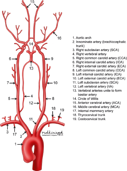
The intracranial arterial system is divided into the anterior and posterior circulations. The anterior circulation includes the internal carotid artery and its branches as well the anterior and middle cerebral arteries and anterior and posterior communicating arteries. The posterior circulation includes the vertebral arteries, basilar artery, and posterior cerebral arteries ( Fig. 5.1 ).

Fig. 5.1 Coronal view of the arteries from the aortic arch, which supply the brain.

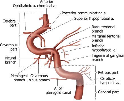
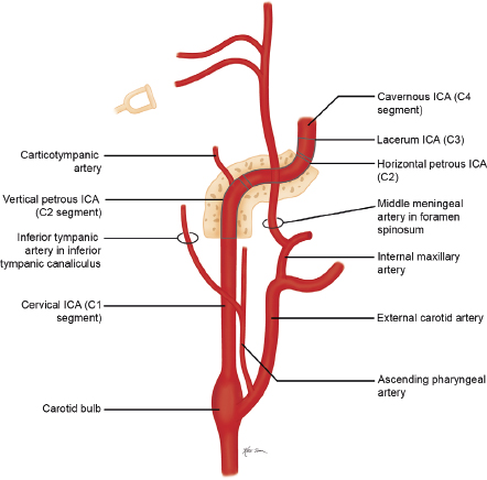
The internal carotid artery (ICA) is divided into seven segments corresponding to their embryonic precursor arteries ( Fig. 5.2 and Fig. 5.3 ). The first ICA segment extends from the bifurcation of the common carotid artery in the neck to the inferior level of the skull base. The second ICA segment is located within the petrous carotid canal of the temporal bone and has two small branches, the vidian artery (artery of the pterygoid canal), which anastomoses with branches of the external carotid artery (ECA), and the caroticotympanic artery to the middle ear. The third ICA segment is a short portion superior to the foramen lacerum that extends from the petrous apex to the cavernous sinus. The fourth ICA segment is located within the cavernous sinus and has two major branches (the meningohypophyseal trunk, which supplies the pituitary gland; clival dura; and tentorium via basal and marginal tentorial branch arteries, inferior hypophyseal artery, and trigeminal ganglion artery; and the inferolateral trunk, which supplies the cranial nerves and dura of the cavernous sinuses via cavernous sinus and meningeal branch arteries), which have anastomoses with branches of the ECA. The fifth ICA segment courses within the anterior portion of the cavernous sinus until it exits superiorly into the cranial cavity. The ophthalmic artery may occasionally arise from the fifth segment. The sixth ICA segment is the first segment in the intracranial subarachnoid space and has two branches (the ophthalmic artery and the superior hypophyseal artery, which supplies the adenohypophysis, pituitary stalk, and optic chiasm). The seventh ICA segment extends superiorly to the bifurcation of the ICA into the anterior cerebral artery (ACA) and middle cerebral artery (MCA) and includes the posterior communicating artery (PCOM) as a branch.

Fig. 5.2 Lateral view of the segments of the internal carotid artery and its branches. From THIEME Atlas of Anatomy, Neck and Internal Organs, © Thieme 2006, Illustration by Karl Wesker.
Fig. 5.3 Coronal view of the segments of the internal carotid artery and its branches.

The ACA has three segments ( Fig. 5.4 and Fig. 5.5 ). The A1 segment extends medially from the ICA to the midline and the origin of the anterior communicating artery (ACOM), which connects to the contralateral ACA. The A1 segment has branch arteries, such as the medial lenticulostriate arteries, that provide blood supply to the basal ganglia. The A2 segment extends superiorly within the interhemispheric fissure from the ACOM to the level of the rostrum of the corpus callosum. Branch arteries from the A2 segment include the orbitofrontal and frontopolar arteries, which supply the inferomedial portions of the frontal lobes. The recurrent artery of Heubner can also arise from the proximal portion of A2 providing blood to the basal ganglia in addition to the lenticulostriate arteries from the A1 segment. In some cases, the recurrent artery of Heubner may arise from the A1 segment or anterior communicating artery. The A3 segment extends around the corpus callosum and divides into the pericallosal and callosomarginal arteries, which course posteriorly over the corpus callosum and cingulate gyrus, respectively. The A2 and A3 segments provide blood to the medial portions of the frontal and parietal lobes, corpus callosum, and anterior limbs of the internal capsules.

Fig. 5.4 Basal view of the anterior and middle cerebral arteries, basilar artery, and posterior cerebral arteries and their branches.
Fig. 5.5 Sagittal view of the anterior cerebral arteries and their branches. From THIEME Atlas of Anatomy, Neck and Internal Organs, © Thieme 2006, Illustration by Karl Wesker.

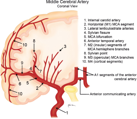
The MCA has four segments ( Fig. 5.6, Fig. 5.7 , and Fig. 5.8 ). The M1 (horizontal) segment extends laterally from the ICA to the sylvian fissure and has branch arteries, including the lateral lenticulostriate arteries, which supply the external capsule, caudate nucleus, and putamen, and the anterior temporal artery, which supplies the anterior portions of the temporal lobes. At the level of the sylvian fissure, the M1 segment bifurcates into the M2 (insular) segments, which extend upward and posteriorly along the insula. Branches from the M2 segments extend laterally along the overhanging or opercular portions of the cerebral hemispheres (M3 segments), providing blood to these locations. Continuation of the M3 segments after they exit the sylvian fissure are the M4 (cortical) segments, which supply the lateral portions of the cerebral hemispheres.

Fig. 5.6 Coronal view of the middle cerebral arteries and their branches.
Fig. 5.7 Lateral surface view of the middle cerebral arteries and their superficial branches. From THIEME Atlas of Anatomy, Neck and Internal Organs, © Thieme 2006, Illustration by Karl Wesker.
Fig. 5.8 Lateral view within the sylvian fissure shows the middle cerebral arteries and their proximal branches. From THIEME Atlas of Anatomy, Neck and Internal Organs, © Thieme 2006, Illustration by Karl Wesker.

The upper terminal branches of the basilar artery are the posterior cerebral arteries (PCAs), which supply blood to the posteroinferior portions of the parietal lobes, occipital lobes, and thalami ( Fig. 5.9 and Fig. 5.10 ). The PCAs have four segments. The P1 segment extends laterally from the basilar artery to the level of the posterior communicating artery (PCOM). Branches from the P1 segment include the posterior thalamoperforating arteries, which supply the midbain and posterior portions of the thalami. The P2 segment extends posterolaterally from the P1 segment– PCOM junction around the midbrain above the tentorium. Arterial branches from the P2 segment include the anterior and posterior temporal arteries, which provide blood to the inferior portions of the temporal lobes not supplied by the anterior temporal artery from the M1 segment of the MCA. Other branches from the P2 segment include the medial and lateral posterior choroidal arteries, which supply the choroid plexus of the third and lateral ventricles, respectively; the thalamogeniculate arteries, which supply the posterior thalamus; and the peduncular perforating arteries, which supply the midbrain. The P3 segment is the portion of the PCA located in the quadrigeminal cistern posterior to the midbrain prior to its entry into the calcarine fissure. The P4 segment of the PCA is located within the calcarine fissure and its branches include the parieto-occipital artery, calcarine artery, lateral occipital artery, and posterior splenial arteries, which supply the occipital lobe, posterior medial portions of the temporal lobes, and posterior corpus callosum.

Fig. 5.9 Basilar view of the posterior cerebral artery and its branches.
Fig. 5.10 Lateral view of the posterior cerebral artery and its branches.

Single vertebral arteries (VAs) arise from each subclavian artery and extend superiorly in the neck to eventually fuse intracranially to form the basilar artery anterior to the brainstem near the pontomedullary junction ( Fig. 5.1 ). Each vertebral artery is divided into four segments. The V1 segment of the VA extends from its origin from the subclavian artery to its entry into the C6 foramen transversarium. The V1 segment supplies the lower portion of the cervical spinal cord and paraspinal musculature. The V2 segment extends upward through the C6 to C1 foramina tranversaria. The anterior meningeal artery is a branch from the V2 segment. The V3 segment is the portion of the VA from its exit from the C1 transverse foramen to the level of the outer dural margin at the foramen magnum. The posterior meningeal artery is a branch from the V3 segment. The V4 segment is the intradural portion of the VA from which the posterior inferior cerebellar artery arises. Other branches from the V4 segment include medullary perforating arteries and anterior and posterior spinal arteries. Both V4 segments fuse to form the basilar artery. Branches from the basilar artery include the anterior inferior cerebellar artery, superior cerebellar arteries, and basilar perforating arteries ( Fig. 5.11 and Fig. 5.12 ). The uppermost portion of the basilar artery terminates with a bifurcation into the right and left posterior cerebral arteries. The upper vertebral arteries and basilar artery provide blood supply to the upper spinal cord, brainstem, and cerebellum.

Fig. 5.11 Lateral view of the upper vertebral and basilar artery and their branches. From THIEME Atlas of Anatomy, Neck and Internal Organs, © Thieme 2006, Illustration by Karl Wesker.
Fig. 5.12 Lateral view of the upper vertebral and basilar artery and their cerebellar branches.

The circle of Willis is an important intracranial anastomotic arterial ring around the lower brain interconnecting the anterior and posterior circulations and includes the upper portions of both ICAs, the A1 segments of the ACAs, the anterior communicating artery (ACOM), the basilar artery, posterior communicating arteries (PCOMs), and P1 segments of both posterior cerebral arteries (PCA).


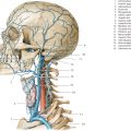
The external carotid artery (ECA) arises from the bifurcation of the cervical portion of the common carotid artery. The ECA has eight major branches ( Fig. 5.13 ). The superior thyroid artery extends inferiorly to supply the larynx and thyroid gland. The ascending pharyngeal artery extends superiorly to supply the nasopharynx, oropharynx, dura, middle ear, and cranial nerves IX, X, and XI. The lingual artery supplies the oral cavity, tongue, and submandibular gland. The facial artery supplies the face, cheek, lips, and palate. The occipital artery supplies the meninges of the posterior cranial fossa as well as the upper cervical paraspinal muscles and scalp. The posterior auricular artery supplies the scalp and outer ear. The superficial temporal artery supplies the scalp. The maxillary artery supplies the deep soft tissues of the face and nose. A branch from the maxillary artery extends superiorly through the foramen spinosum as the middle meningeal artery to supply the intracranial meninges. Anastomoses between ECA branches (except for the superior thyroid and lingual arteries) and intracranial branches of the ICA and/or vertebral arteries can occur.

Fig. 5.13 Oblique coronal view of the external carotid artery and its branches.


Intracranial Venous Anatomy


The intracranial venous system consists of a superficial system that drains blood from the cerebral cortex and superficial white matter into cortical veins and eventually into the dural venous sinuses, and a deep venous system that drains blood from the deep white matter and basal ganglia ( Fig. 5.14 , Fig. 5.15 , and Fig. 5.16 ). The deep venous system includes the paired internal cerebral veins in the roof of the third ventricle, the basal vein of Rosenthal, the vein of Galen, and the straight venous sinus. Also included in the deep venous system are transcerebral veins. The internal cerebral veins represent the confluence of septal, subependymal, ventricular, anterior caudate, thalomostriate, and choroidal veins at the foramen of Monro. The basal veins of Rosenthal are located at the medial aspects of the temporal lobes and drain blood from the adjacent temporal lobes, insula, and cerebral peduncles. These veins have anastomoses with the middle cerebral veins and petrosal veins. The basal veins course posteriorly around the cerebral peduncles, where they unite with internal cerebral veins to form the single midline great vein of Galen located below the splenium of the corpus callosum. The vein of Galen connects with the inferior sagittal sinus to drain venous blood into the straight venous sinus. Transcerebral veins are medullary veins that extend though the cerebral tissue and are not usually visualized on normal MRI or CT examinations.

Fig. 5.14 Lateral view of the superficial and deep cerebral venous systems. From THIEME Atlas of Anatomy, Neck and Internal Organs, © Thieme 2006, Illustration by Karl Wesker.
Fig. 5.15 Medial view of the superficial and deep cerebral venous systems. From THIEME Atlas of Anatomy, Neck and Internal Organs, © Thieme 2006, Illustration by Karl Wesker.
Fig. 5.16 Inferior basal view of the superficial and deep cerebral venous systems. From THIEME Atlas of Anatomy, Neck and Internal Organs, © Thieme 2006, Illustration by Karl Wesker.

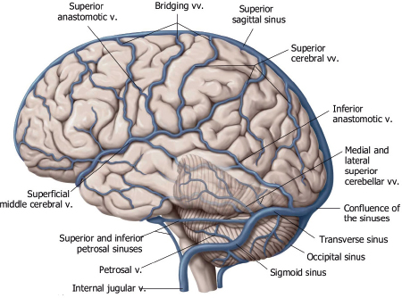
The superficial venous system includes superficial cerebral veins along the surface of the brain that drain blood from the adjacent cerebral cortex and subcortical white matter. The superficial veins can have various configurations, including approximately 12 that drain into the superior sagittal sinus. A prominent superficial anastomotic vein of Trolard can be observed that connects to the superior sagittal sinus. A prominent inferior anastomotic superficial vein of Labbé can also be seen that connects the transverse venous sinus. The superficial middle cerebral vein is another prominent vein that courses anteriorly in the sylvian fissure and drains blood from the adjacent lateral brain into the cavernous sinuses and/or pterygoid venous plexus.


Venous Sinuses


Veins from the superficial and deep venous systems drain into the dural venous sinuses ( Fig. 5.17 ). The dural venous sinuses are endothelial channels located between the periosteal (outer) and meningeal (inner) layers of the dura. The dural venous sinuses can be subdivided into two groups: posterosuperior and anteroinferior. The posterosuperior group includes the superior and inferior sagittal sinuses, straight venous sinus, torcular herophili, transverse and sigmoid sinuses, and jugular bulbs. Arachnoid granulations can occur in all of the dural venous sinuses, although they are most frequently present in the superior sagittal and transverse venous sinuses.

Fig. 5.17 Sagittal view of the intracranial venous drainage patterns, including the venous sinuses. From THIEME Atlas of Anatomy, Neck and Internal Organs, © Thieme 2006, Illustration by Markus Voll.

The anteroinferior group of dural venous sinuses includes the cavernous sinus, superior and inferior petrosal sinuses, clival venous sinus, and sphenoparietal sinus. Blood from the cavernous sinuses drain into pterygoid venous plexuses via the foramen ovale or into the superior and/or inferior petrosal sinuses. Blood from the superior petrosal sinus drains into the sigmoid sinus, whereas blood from the inferior petrosal sinus drains into the jugular bulb. The clival venous plexus connects the cavernous sinus with the petrosal sinuses. The sphenoparietal sinus at the anterior portion of the middle cranial fossa connects the superficial veins adjacent to the temporal lobes to the inferior petrosal sinus ( Fig. 5.17 ).


The veins in the posterior cranial fossa can drain upward into the vein of Galen (superior vermian vein, precentral cerebellar vein, and anterior pontomesencephalic vein), anteriorly into the petrosal sinuses (petrosal veins), or posterolaterally into the transverse or sigmoid venous sinuses (inferior vermian veins) ( Fig. 5.18 ).

Fig. 5.18 Inferior basal view of the veins of the brainstem. From THIEME Atlas of Anatomy, Neck and Internal Organs, © Thieme 2006, Illustration by Karl Wesker.


Cerebral Arterial and Venous Development


Arterial Development


There are six branchial arches during embryonic development. Derivatives of the third branchial arches form the internal carotid arteries. A portion of the left fourth branchial arch forms most the aortic arch, and the right fourth branchial arch forms the proximal segment of the right subclavian artery. Derivatives of the first arch form the vidian artery. Derivatives of the second arch form the pharyngeal artery, external carotid artery, stapedial artery, and caroticotympanic artery.


At 4 weeks of gestation, primitive, paired internal carotid arteries connected to the dorsal aorta and third aortic arch provide blood to the developing vesicles of the forebrain, midbrain, and hindbrain. The mature pattern of the cerebral arterial system develops by 8 weeks of gestation.


At 4 weeks of gestation, the developing hindbrain is supplied by paired longitudinal arteries that have transient anastomoses with the internal carotid arteries. These paired arteries progressively fuse in the midline to form the basilar artery around 5 weeks of gestation. Lack of normal fusion of the longitudinal arteries can result in arterial fenestrations. Lack of normal involution of some of the anastomoses between the internal carotid arteries and longitudinal arteries can result in anomalous connections between the internal carotid arteries and basilar artery, such as a persistent trigeminal artery ( Fig. 5.19 ).

Fig. 5.19 Lateral diagram shows potential developmental anomalous connections between the internal carotid arteries and basilar artery.


Venous Development


At the fifth week of gestation, three venous plexuses form around the posterolateral aspects of the developing brain. Progressive growth and connections between these plexuses result in eventual formation of the major dural venous sinuses over the next 6 months.


Development of cortical veins occurs after normal involution of early fetal transcerebral veins, and the primitive pial venous plexus develops by 11 weeks of gestation.


CT Angiography and CT Perfusion


CT angiography (CTA) is a powerful imaging modality for evaluating normal and abnormal blood vessels. CTA has proven to be clinically useful in the evaluation of intracranial arteries, veins, and dural venous sinuses. Pathologic processes involving intracranial blood vessels, such as aneurysms, arteriovenous malformations, arterial occlusions, and dural venous sinus thrombosis, can be seen with CTA.


CT perfusion is a relatively new technique using dynamic intravenous infusion of contrast to measure cerebral blood flow (CBF), cerebral blood volume (CBV), and mean transit time of contrast enhancement (MTT) in selected volumes of interest in the brain. CT perfusion has major clinical application in the evaluation of cerebral infarcts and adjacent zones of decreased perfusion (penumbra and oligemic areas) at risk for progression to infarction. Maintenance of CBF is critical for neuronal function. With arterial occlusion, loss of normal neuronal electrical activity occurs within seconds after arterial occlusion. Cellular death is dependent on the duration and magnitude of ischemia, metabolic vulnerability of specific anatomic sites, and the oxygen content of blood. Normal CBF ranges from 50 to 60 mL/100 g/min. When CBF is reduced to 15 to 20 mL/100 g/min for several hours (mild–moderate hypoxia), spontaneous and evoked neuronal electrical activity decreases significantly secondary to ischemia, although it can be reversed by reperfusion with CBF above 50 mL/100 g/min. With severe hypoxia/anoxia resulting from CBF below 10 mL/100 g/min, cellular membrane depolarization and ischemia leading to brain infarction may occur within several minutes.


When thrombotic or embolic arterial occlusions occur, CBF in the involved brain tissue is usually heterogeneous, with a central core showing the greatest reductions in CBF that cause irreversible cell damage and infarction, and a surrounding zone (referred to as the salvageable penumbra) that may have moderate reduction in CBF, resulting in ischemia that may be reversible with reperfusion. The penumbra typically shows loss of neuronal electrical activity without immediate anoxic depolarization, as well as loss of autoregulation. If reperfusion does not occur, the penumbra will progress to infarction. An oligemic zone of mildly reduced CBF may also be seen surrounding the penumbra, and this zone is less vulnerable to infarction than the penumbra. Thrombolytic medication can be useful and beneficial when it results in timely reperfusion to the penumbra and oligemic zone. Estimating the sizes of the penumbra and oligemic zone can be done in the acute setting with dynamic contrast-enhanced CT. CT perfusion, utilizing iodinated contrast delivered as an intravenous bolus, can use the linear relationship between contrast concentration and attenuation to directly calculate and quantify CBF, CBV, and MTT for sites of ischemia and infarction in the brain prior to thrombolytic treatment.


Magnetic Resonance Angiography


Magnetic resonance imaging (MRI) is a powerful modality for evaluating normal and abnormal blood vessels. The appearance of blood vessels on MRI depends on various factors, such as the type of MRI pulse sequence, pulsatility and range of velocities in the vessels of interest, and size, shape, and orientation of the vessels relative to the image plane. Useful anatomic information about blood vessels can be gained by using spin echo pulse sequences, which can display patent vessels as zones of signal void (blackblood images), or gradient recalled echo (GRE) pulse sequences, which display the moving hydrogen atomic nuclei (protons) in blood as zones of high signal (brightblood images).


The GRE technique is used to generate MR angiograms (MRA). The high signal from flowing blood on GRE images reflects movement patterns and velocities of hydrogen atomic nuclei rather than direct anatomic displays of the blood vessels. The operator of the MRI equipment can choose parameters to optimize the imaging of various arteries and veins. Two main types of GRE techniques are used for MRA. One is based on hydrogen signal amplitude and is referred to as the time-of-flight (TOF) method. The other method is based on the phase differences of the moving protons (hydrogen) in blood compared with stationary tissue and is referred to as phase-contrast (PC) MRA.


In TOF MRA, the GRE pulse sequence is optimized for demonstrating the inflow enhancement (high signal) of moving protons (hydrogen nuclei) in blood relative to the low signal of protons in stationary tissue. Phase-contrast (PC) MRA is a technique that differentiates flowing and stationary protons through the use of bipolar flow-encoding gradients. If the flow velocity is known, the flow sensitivity of the sequence can be selected to emphasize the vessels of interest. PC MRA can be optimized for detecting slow flow in veins and at areas of high-grade arterial stenosis.


The individual GRE images can be acquired in a sequential mode, also referred to as two-dimensional (2D) TOF or PC MRA, or as an entire volume of covered tissue, which is referred to as three-dimensional (3D) TOF or PC MRA. The acquired image data from either of these two methods are post-processed with computer algorithms to generate the MRA images in a display format similar to conventional arteriograms. Two commercially available types of post-processing are the maximum intensity projection (MIP) technique and surface rendering (SR). The former technique is more common, and the MIP MRA images can be displayed in any plane of obliquity on film or as a movie loop. Surface rendering is another post-processing method for MRA that shows 3D relationships by giving the displayed vessels shadowing and perspective. The MRA images are projected in a fashion similar to that used for the MIP method. Surface rendering has been demonstrated to be useful in showing spatial relationships between vessels on a single coronal image, allowing differentiation of adjacent and overlapping vessels.


MRA has proven to be clinically useful in the evaluation of the carotid arteries in the neck, intracranial arteries, intracranial veins, and dural venous sinuses. Disorders like aneurysms, arteriovenous malformations, arterial occlusions, dural venous sinus thrombosis, etc., can be seen with MRA.


References

1. Johnson MH, Thorisson HM, Diluna ML. Vascular anatomy: the head, neck, and skull base. Neurosurg Clin N Am 2009;20(3):239–258 2. Kathuria S, Chen J, Gregg L, Parmar HA, Gandhi D. Congenital arterial and venous anomalies of the brain and skull base. Neuroimaging Clin N Am 2011;21(3):545–562, vii 3. Kathuria S, Gregg L, Chen J, Gandhi D. Normal cerebral arterial development and variations. Semin Ultrasound CT MR 2011;32(3):242–251 4. Raybaud C. Normal and abnormal embryology and development of the intracranial vascular system. Neurosurg Clin N Am 2010;21(3):399–426 5. Scott JN, Farb RI. Imaging and anatomy of the normal intracranial venous system. Neuroimaging Clin N Am 2003;13(1):1–12


Table 5.1 Congenital and developmental vascular anomalies/variants


  • Persistent fetal origin of posterior cerebral artery


  • Hypoplasia of the A1 segment of the anterior cerebral artery


  • Persistent trigeminal artery (PTA)


  • Persistent otic artery


  • Persistent hypoglossal artery


  • Proatlantal artery


  • Duplications of cerebral, carotid, vertebral, or basilar arteries


  • Azygous anterior cerebral artery


  • Hemiazygous anterior cerebral artery


  • Arterial fenestration


  • Aberrant position of the internal carotid artery


  • Persistent stapedial artery (PSA)


  • Unilateral agenesis, aplasia, and hypoplasia of the internal carotid artery


  • Vein of Galen aneurysm


  • Sturge-Weber syndrome


  • Moyamoya disease


  • ACTA2 mutations with dolichoectasia of the proximal internal carotid arteries and stenosis of the upper internal carotid arteries


  • Menkes’ syndrome


  • PHACES syndrome (posterior fossa malformations, facial hemangiomas, arterial anomalies, cardiac anomalies and aortic coarctation, eye abnormalities, and sternal clefts or supraumbilical raphe)


  • Thoracic outlet syndrome


  • Venous angioma (developmental venous anomaly)


  • Dehiscence of the jugular bulb


  • High position of the jugular bulb


  • Sinus pericranii


Table 5.1 Congenital and developmental vascular anomalies/variants

Lesion


Imaging Findings


Comments


Persistent fetal origin of posterior cerebral artery ( Fig. 5.20 )


Large posterior communicating artery supplying the posterior cerebral artery, associated with hypoplasia or absence of connection between the basilar artery and the ipsilateral posterior cerebral artery.


Represents persistence of embryonic configuration, common vascular variant seen in ~ 20% of arteriograms.


Hypoplasia of the A1 segment of the anterior cerebral artery ( Fig. 5.21 )


Hypoplasia or absent A1 segment associated with a patent communicating artery supplying blood to ipsilateral A2 segment.


Anatomic variant seen in ~ 10% of arteriograms.


Persistent trigeminal artery (PTA) ( Fig. 5.22 and Fig. 5.23 )


Anomalous anastomosis connecting the internal carotid artery in cavernous sinus to the basilar artery at the level of the trigeminal nerve. Can pass posteriorly either lateral or medial to the sella turcica. The medially positioned PTA can indent the pituitary gland and needs to be reported if surgery is planned. The basilar artery below the anastomosis and the vertebral arteries are usually small.


Most common type of anomalous carotid-basilar anastomosis (0.5% of cerebral arteriograms), caused by failure of involution of persistent embryonic circulatory configuration. Associated with increased incidence of aneurysms and vascular malformations. Other less common types of anomalous carotidbasilar anastomosis include: persistent hypoglossal artery (adjacent to CN XII), persistent otic artery, and proatlantal intersegment artery.


Persistent otic artery


Anomalous anastomosis connecting the petrous portion of the internal carotid artery medially through the internal auditory canal to the lower basilar artery.


Rarest of anomalous carotid-basilar anastomoses, caused by failure of involution of persistent embryonic circulatory configuration. Associated with increased incidence of aneurysms and vascular malformations.


Persistent hypoglossal artery ( Fig. 5.24 )


Anomalous anastomosis connecting the posterior upper cervical portion of the internal carotid artery at the C1–C2 level to the basilar artery along the hypoglossal nerve within an enlarged hypoglossal canal. Often provides all of the blood flow to the basilar artery. Vertebral arteries are usually small.


Anomalous carotid-basilar anastomosis (0.1% of cerebral arteriograms), caused by failure of involution of persistent embryonic circulatory configuration. Associated with increased incidence of aneurysms and vascular malformations.


Proatlantal artery


Anomalous anastomosis connecting the posterior cervical portion of the internal carotid artery at the C2–C3 level to the suboccipital vertebral artery prior to intracranial entry through the foramen magnum.


Failure of involution of persistent embryonic circulatory configuration. Associated with increased incidence of aneurysms and vascular malformations.


Duplications of cerebral, carotid, vertebral, or basilar arteries ( Fig. 5.25 and Fig. 5.26 )


Duplication of arteries usually occurs as two parallel arteries from two separate origins, as seen on CTA, MRA, and/or conventional angiography.


Duplicated arteries have two origins and variable courses with or without eventual fusion. Duplications of intracranial or cervical arteries are less frequent than other anomalies of intracranial arteries. Other less common variants include fenestrations and accessory arteries.


Azygous anterior cerebral artery


Solitary A2 branch distal to the A1 segments of the anterior cerebral arteries.


Developmental variant with one A2 segment present distal to the A1 segments. Associated with holoprosencephaly.


Hemiazygous anterior cerebral artery ( Fig. 5.27 )


Both A2 segments of the anterior cerebral arteries arise from a more proximal solitary artery distal to the A1 segments.


Developmental variant with two A2 segments of the anterior cerebral arteries arising from a solitary proximal trunk.


Arterial fenestration ( Fig. 5.28 )


Duplication of a portion of an artery whose main trunk is derived from a single origin, as seen on CTA, MRA, or conventional angiography.


Developmental variation when there are double segments involving portions of the vertebral, basilar, or carotid arteries. With arterial fenestration, a vessel with a single origin divides into two parallel segments along its course with eventual re-anastomosis.


Aberrant position of the internal carotid artery ( Fig. 5.29 )


Abnormal position of the internal carotid artery (ICA), which enters the middle ear posteriorly through an enlarged inferior tympanic canaliculus lateral to the expected site of the petrous carotid canal. The anomalous artery courses anteriorly over the cochlear promontory to connect with the horizontal petrous ICA via a dehiscent carotid bone plate. The aberrant ICA in the middle ear is usually smaller in caliber than the contralateral normal ICA.


Congenital arterial variation related to altered formation of the extracranial ICA resulting from agenesis of the normal first embryonic segment of the ICA. A collateral alternative developmental pathway occurs where the proximal ICA originates from the ascending pharyngeal artery connecting to the inferior tympanic artery, which extends superiorly through the inferior tympanic canal into the middle ear where it anastomoses with the caroticotympanic artery, which connects to the lateral petrous portion of the ICA. As a result, the ICA is positioned laterally within the middle ear cavity. Also, a characteristic narrowing of the inferior tympanic artery occurs as it passes through the inferior tympanic canal at the skull base. Often an incidental finding with surgical planning implications.


Persistent stapedial artery (PSA) ( Fig. 5.30 )


Commonly occurs as an anomalous small artery associated with an aberrant internal carotid artery (ICA) or as an isolated anomaly. Findings include an absent ipsilateral foramen spinosum. The small tubular PSA extends from the ICA along the cochlear promontory through the stapes and then adjacent to the tympanic segment of CN VII to an enlarged tympanic facial nerve canal, where it enters the middle cranial fossa as the middle meningeal artery.


Rare vascular anomaly that is often associated with an aberrant ICA. Results from lack of normal evolution of the embryonic hyoid artery (from the second anterior embryonic aortic arch) into the stapedial artery, with eventual formation of the branches of the external carotid arteries (ECA) supplying the orbits, meninges, and lower face, as well as the small caroticotympanic and superior tympanic arteries. Lack of normal involution of the stapedial artery results in a persistent stapedial artery that extends from the ICA into the middle ear, passing through the stapes near the course of the tympanic portion of CN VII and extending intracranially to supply the middle meningeal artery. As a result, the middle meningeal artery is not supplied by the ECA and internal maxillary artery. There is no ipsilateral foramen spinosum.


Unilateral agenesis, aplasia, and hypoplasia of the internal carotid artery ( Fig. 5.31 )


Absence or near-complete absence of the internal carotid artery (ICA) and petrous carotid canal.


Congenital arterial variation that occurs in less than 0.01% of the population. Results from abnormal embryonic development of the third aortic arch and dorsal aorta from which the internal carotid artery arises. Collateral intracranial blood flow occurs via patent anterior and/or posterior communicating arteries.


Vein of Galen aneurysm ( Fig. 5.32 )


Multiple, tortuous, contrast-enhancing vessels involving choroidal and thalamoperforate arteries, internal cerebral veins, vein of Galen (aneurysmal formation), straight and transverse venous sinuses, and other adjacent veins and arteries. The venous portions often show contrast enhancement. CTA shows contrast enhancement in patent portions of the vascular malformation.


Heterogeneous group of vascular malformations with arteriovenous shunts and dilated deep venous structures draining into and from an enlarged vein of Galen, ± hydrocephalus, ± hemorrhage, ± macrocephaly, ± parenchymal vascular malformation components, ± seizures and high-output congestive heart failure in neonates.


Sturge-Weber syndrome ( Fig. 5.33 and Fig. 5.34 )


Prominent, localized, unilateral leptomeningeal contrast enhancement usually in parietal and/or occipital regions in children, ± gyral enhancement, mild localized atrophic changes in brain adjacent to the pial angioma, ± prominent medullary and/or subependymal veins, ± ipsilateral prominence of choroid plexus. Gyral calcifications > 2 years, with progressive cerebral atrophy in region of pial angioma.


Also known as encephalotrigeminal angiomatosis, Sturge-Weber syndrome is a neurocutaneous syndrome associated with ipsilateral port wine cutaneous lesion and seizures. It results from persistence of primitive leptomeningeal venous drainage (pial angioma) and developmental lack of normal cortical veins, producing chronic venous congestion and ischemia.


Moyamoya disease ( Fig. 5.35 )


Multiple, tortuous, small, enhancing vessels may be seen in the basal ganglia and thalami secondary to dilated collateral arteries, + enhancement of these arteries related to slow flow within the collateral arteries versus normal-sized arteries. Contrast enhancement of the leptomeninges related to pial collateral vessels. Decreased or absent contrast enhancement in the supraclinoid portions of the internal carotid arteries and proximal middle and anterior cerebral arteries.


MRA and CTA show stenosis and occlusion of the distal internal carotid arteries with collateral arteries (lenticulostriate, thalamoperforate, and leptomeningeal) best seen after contrast administration enabling detection of slow blood flow.


Progressive occlusive disease of the intracranial portions of the internal carotid arteries, with resultant numerous dilated collateral arteries arising from the lenticulostriate and thalamoperforate arteries as well as other parenchymal, leptomeningeal, and transdural arterial anastomoses. Moyamoya means “puff of smoke,” referring to the angiographic appearance of the collateral arteries (lenticulostriate and thalamoperforate). The disease usually has a nonspecific etiology but can be associated with neurofibromatosis, radiation angiopathy, atherosclerosis, sickle-cell disease, and mutations in BRCC3/MTCP1 and GUCY1A3 genes. Usually occurs in children more than in adults, and in Asia more than other locations.


ACTA2 mutations with dolichoectasia of the proximal internal carotid arteries and stenosis of the upper internal carotid arteries ( Fig. 5.36 )


Conventional arteriography, MRA, CTA: Dilatation of the proximal internal carotid arteries (ICAs), severe narrowing of the upper ICAs, straightened patterns of the proximal ICAs, stenosis or occlusion of the M1 segments of the middle cerebral arteries without lenticulostriate collaterals, ± tortuous or corkscrew appearance of distal anterior, middle, and posterior cerebral arteries.


MRI: Multiple small cerebral infarcts involving the cerebral cortex and/or white matter.


Arg179 missense mutation of the ACTA2 gene is associated with smooth muscle dysfunction resulting in dilatation of the proximal ICAs, severe narrowing of the upper ICAs, straightened patterns of the proximal ICAs, “moyamoya-type” collateral vessels, large artery occlusions, distal small distal aneurysms, dilated extradural arteries; as well as patent ductus arteriosus, mydriasis, pulmonary hypertension, and gastrointestinal and bowel dysfunction. Usual presentation is in children. ACTA2 mutations result in abnormal fibromuscular and/or smooth muscle proliferation in the intima and media of arteries.


Menkes’ syndrome ( Fig. 5.37 )


MRI: High signal on T2-weighted imaging can be seen in the cerebral white matter, putamen bilaterally, and/or caudate nuclei, with or without restricted diffusion. Delayed myelination involving the posterior limbs of the internal capsules can be seen. Progressive atrophy of the cerebrum, cerebellum, and brainstem. Small zones with high signal on T1-weighted imaging may be seen in the cerebral cortex. Large bilateral subdural hematomas can be seen with mixed signal on T1- and T2-weighted imaging.


MRA and CTA show tortuous “corkscrew” arteries.


CT: Rapid, progressive brain atrophy with large bilateral subdural hematomas.


X-linked recessive disorder from mutations of the ATP7A gene on Xq13.3, which encodes for the copper-transporting ATPase necessary for intestinal uptake of copper. Lack of adequate copper results in defective cytochrome c oxidase activity in mitochondria. Patients often have seizures, truncal hypotonia, hypothermia, failure to thrive, lack of reaching developmental milestones, hypermobile joints, hypopigmentation, and coarse, stiff, and broken hair—“kinky hair disease.”


PHACES syndrome (posterior fossa malformations, facial hemangiomas, arterial anomalies, cardiac anomalies and aortic coarctation, eye abnormalities, and sternal clefts or supraumbilical raphe)( Fig. 5.38 )


Vascular anomalies occur in 30% of patients. Arterial abnormalities include: absence or hypoplasia of the carotid, vertebral, and/or cerebral arteries; anomalous arteries; arterial stenoses and occlusions; moyamoya; aneurysms; and AVMs. Cavernous malformations can also occur. Malformations in the posterior cranial fossa can include Dandy-Walker malformation and dysgenesis/hypoplasia of the cerebellum, corpus callosum, or septum pellucidum.


Rare spectrum of anomalies of unknown etiology that includes facial hemangiomas that are present at birth and involve one trigeminal nerve division, as well as one or more of the following: arterial anomalies, coarctation of the aorta, malformations in the posterior cranial fossa, eye abnormalities (microphthalmos, cataracts, iris hypoplasia, and optic nerve hypoplasia or atrophy), cardiac defects, and/or sternal clefts. Occurs predominantly in females (up to 90% of cases).


Thoracic outlet syndrome ( Fig. 5.39 )


Cervical ribs or fibrous bands located adjacent to the subclavian artery, subclavian vein, and/or brachial plexus.


Signs and symptoms of the thoracic outlet syndrome (TOS) occur from compression of the brachial plexus (neurogenic TOS), subclavian artery (arterial TOS), and/or subclavian vein (venous TOS). Neurogenic TOS accounts for ~ 90% of cases. Compression of the thoracic outlet structures can be static or positional. Causes of the compression include cervical ribs, fibrous bands, or hypertrophy or anomalies of the scalene muscles.


Venous angioma (developmental venous anomaly) ( Fig. 5.40 and Fig. 5.41 )


MRI: Gadolinium contrast enhancement of one or two prominent intra-axial vein(s) connected to a network of multiple small draining veins (caput medusae). Often no findings on T1- or T2-weighted imaging unless the vein is prominent. Some developmental venous anomalies have slightly high signal on T2-weighted imaging. Low signal on SWI can be seen at these venous anomalies.


CT: No abnormality or small, slightly hyperdense zone prior to contrast administration. Contrast enhancement seen in a slightly prominent vein draining a collection of small veins on contrast-enhanced CT and CTA.


Considered an anomalous venous formation. Typically not associated with hemorrhage. Usually an incidental finding, except when it is associated with cavernous malformation (in ~ 25% of cases).


Dehiscence of the jugular bulb ( Fig. 5.42 )


Protrusion of the jugular bulb into the posteroinferior portion of the middle ear related to deficient or absent bone at the jugular plate.


Venous variant anatomy with the jugular bulb extending superiorly and laterally into the middle ear through localized bone deficiency/dehiscence of the jugular plate. May be associated with pulsatile tinnitus, Ménière disease, and hearing loss. Important to report for surgical planning.


High position of the jugular bulb


The upper portion of the jugular bulb is located above the base of the internal auditory canal/basilar turn of the cochlea. Does not protrude into the middle ear.


Developmental venous variant anatomy, with positioning of the upper portion of the jugular bulb above the level of the base of the internal auditory canal. Usually an incidental finding.


Sinus pericranii ( Fig. 5.43 )


Communication between dilated extracranial veins and the intracranial veins or dural venous sinuses through a skull defect or emissary veins.


Lesions are nonpulsatile, asymptomatic, soft tissue masses in the scalp near the midline calvarial sutures and often measure 15 mm. Can increase in size with Valsalva maneuver. Lesions are associated with intracranial anomalies, such as solitary DVAs (eight of 13 patients), vein of Galen hypoplasia (two of 13 patients), vein of Galen aneurysm (one of 13 patients), dural sinus malformation (one of 13 patients), and intraosseous arteriovenous malforamtion (one of 13 patients). Can be the cutaneous sign of an underlying venous anomaly.

Fig. 5.20 Persistent fetal origin of posterior cerebral artery. Axial MRA shows the right posterior cerebral artery (arrow) receiving its blood flow directly from the right internal carotid artery via a large right posterior communicating artery with an absent P1 segment of the right posterior cerebral artery.
Fig. 5.21 Hypoplasia of the A1 segment of the anterior cerebral artery. Coronal MRA shows absent A1 segment of the left anterior cerebral artery. The A2 segment of the left anterior cerebral artery receives its blood supply via the anterior communicating artery.
Fig. 5.22 Persistent trigeminal artery. Lateral conventional arteriogram shows a persistent trigeminal artery (arrow), which is an arterial anastomosis between the internal carotid artery at the posteroinferior portion of the cavernous sinus and the basilar artery.
Fig. 5.23 Persistent trigeminal artery. (a) Coronal and (b) axial MRA show a persistent right trigeminal artery (arrows) supplying most of the blood to the basilar artery. The basilar artery below the trigeminal artery and both vertebral arteries are small in caliber.
Fig. 5.24 Persistent hypoglossal artery. (a) Coronal MRA shows an anomalous anastomotic artery (arrow) arising from the posterior upper cervical portion of the internal carotid artery at the C1–C2 level, which connects to the basilar artery via an enlarged hypoglossal canal, as seen on (b) axial gradient recalled echo (GRE) imaging (arrow). Vertebral arteries are small.
Fig. 5.25 Duplications of middle cerebral arteries. Axial MRA shows duplications of both middle cerebral arteries (arrows).
Fig. 5.26 Duplication of right anterior cerebral artery. Axial MRA shows duplication of the A1 segment of the right anterior cerebral artery.
Fig. 5.27 Hemiazygous artery. Coronal MRA shows both A2 segments of the anterior cerebral arteries arising from a more proximal solitary artery (arrow) distal to the A1 segments.
Fig. 5.28 Arterial fenestration. Coronal CTA shows a localized duplication and re-anastomosis of the basilar artery (arrow) representing an arterial fenestration.
Fig. 5.29 Aberrant position of the internal carotid artery. Axial CT image shows the right internal carotid artery passing through the middle ear and positioned lateral to the basal turn of the cochlea (arrow).
Fig. 5.30 Persistent stapedial artery (PSA) and aberrant position of the left internal carotid artery. (a) Axial CT image shows abnormal lateral position of the left internal carotid artery within the middle ear (arrow); the artery enters the posterior portion of the middle ear. (b) The PSA is seen as a small branch arising from the upper portion of the aberrant internal carotid artery (arrow) on coronal CT.
Fig. 5.31 Unilateral agenesis of the left internal carotid artery. The left internal carotid artery is completely absent (a) at the level of the jugular foramen on axial CT and (b) at the level of the cochlea on coronal CT.
Fig. 5.32 A 2-day-old female with a vein of Galen aneurysm/malformation that is seen as abnormal enlargement of the flow voids of the basal veins, vein of Galen, straight venous sinus, and torcular herophili on (a) sagittal T1-weighted imaging and (b) axial T2-weighted imaging, with corresponding high venous flow signal on (c) sagittal 2D phase-contrast MRA.
Fig. 5.33 (a) A 15-year-old male with Sturge-Weber syndrome who has gadolinium contrast enhancement in the leptomeninges on the left from the persistent fetal pial angioma (arrows) on axial T1-weighted imaging, as well as (b) an enhancing transmantle (medullary) vein that connects to the enlarged choroid plexus in the atrium of the left lateral ventricle on axial T1-weighted imaging.
Fig. 5.34 (a) A 17-year-old male with an uncommon form of Sturge-Weber syndrome that is seen as flow voids along the ependymal lining of the lateral ventricles and within the cerebral white matter on axial T2-weighted imaging, with (b) corresponding gadolinium contrast enhancement on coronal T1-weighted imaging.
Fig. 5.35 A 3-year-old female with moyamoya disease. Postcontrast axial MRA shows stenosis and occlusion of the distal internal carotid arteries, with gadolinium contrast enhancement of many small, collateral lenticulostriate, thalamoperforate, and leptomeningeal arteries.
Fig. 5.36 A 14-year-old male with ACTA2 mutations and dolichoectasia of the proximal internal carotid arteries, with severe narrowing of the upper internal carotid arteries and M1 segments of the middle cerebral arteries and straightened patterns of proximal intracranial arteries, as seen on (a) coronal MRA and (b) conventional arteriogram. Tortuous corkscrew distal branches of the anterior and middle cerebral arteries are also seen (b). (c) Axial FLAIR shows multiple intra-axial zones with high signal in the cerebral white matter related to small-vessel ischemia.
Fig. 5.37 (a) An 8-month-old female with Menkes’ syndrome with bilateral complex subdural hematomas on axial CT. (b) Coronal MRA shows tortuous internal carotid arteries (arrows) that have a “corkscrew” appearance.
Fig. 5.38 (a) Patient with PHACES syndrome who has a superficial hemangioma involving the scalp at the top of the head on sagittal fat-suppressed T2-weighted imaging (arrows). (b,c) Coronal MRA shows occlusion of the cervical portion of the right internal carotid artery and anomalous arterial connection from the basilar artery to the right middle cerebral artery branches.
Fig. 5.39 Thoracic outlet syndrome. (a) Coronal CT shows bilateral cervical ribs at the C7 vertebra (arrow) that (b) impress on the subclavian arteries bilaterally (arrows) on coronal CTA.
Fig. 5.40 A contrast-enhancing venous angioma (developmental venous anomaly) is seen in the anterior right frontal lobe on (a) axial CTA and (b) coronal T1-weighted imaging.
Fig. 5.41 A contrast-enhancing venous angioma (developmental venous anomaly) is seen in the right cerebellar hemisphere on (a) axial T1-weighted imaging and has high signal on (b) axial T2-weighted imaging.
Fig. 5.42 Coronal CT shows dehiscence of the right jugular bulb (arrow), which protrudes into the right middle ear.
Fig. 5.43 A 4-week-old female with sinus pericranii, which is seen as communication between extracranial veins and the anterior portion of the superior sagittal sinus through a skull defect, as seen as on (a) axial T1-weighted imaging (arrow) and (b) postcontrast T1-weighted imaging (arrow).

Only gold members can continue reading. Log In or Register to continue

Stay updated, free articles. Join our Telegram channel

May 28, 2020 | Posted by in NEUROLOGICAL IMAGING | Comments Off on 5 Vascular Abnormalities

Full access? Get Clinical Tree

Get Clinical Tree app for offline access