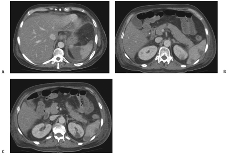
CASE 50 A 42-year-old man with known aortic valve endocarditis presents with left upper abdominal pain and low-grade fever. Fig. 50.1 (A–C) Axial contrast-enhanced CT images show multiple, well-defined, wedge-shaped areas of low attenuation in the spleen and left kidney associated with a left pleural effusion. Contrast-enhanced axial computed tomography (CT) scans show multiple wedge-shaped areas of low attenuation seen in the spleen and the left kidney, along with a small left pleural effusion (Fig. 50.1). Septic emboli causing infarcts in the spleen and left kidney
Clinical Presentation
Radiologic Findings
Diagnosis
Differential Diagnosis
Discussion
Related posts:


Stay updated, free articles. Join our Telegram channel

Full access? Get Clinical Tree


