Case 51
 Clinical Presentation
A 42-year-old woman with increasing abdominal girth.
 Imaging Findings
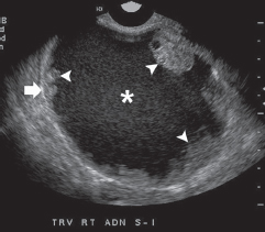
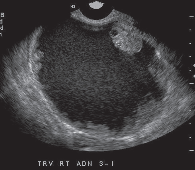
Transvaginal ultrasound images show a complex cystic mass (asterisk) in the right adnexa. The cyst has a thick wall (arrow). Although there are no septa, papillary projections are seen extending into the lumen from the wall of the mass (arrowheads).
 Differential Diagnosis
• Malignant cystic ovarian neoplasm: A thick-walled cystic mass with papillary projections arising from the wall is characteristic.
• Benign ovarian neoplasm: Benign ovarian neoplasms can be large. However, thick walls and papillary projections go against this diagnosis.
• Endometrioma:
Stay updated, free articles. Join our Telegram channel

Full access? Get Clinical Tree


