Case 52
 Clinical Presentation
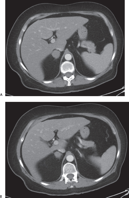
A 41-year-old man with leukemia presents with the sudden onset of right flank pain. Stone protocol computed tomography was performed.
 Imaging Findings
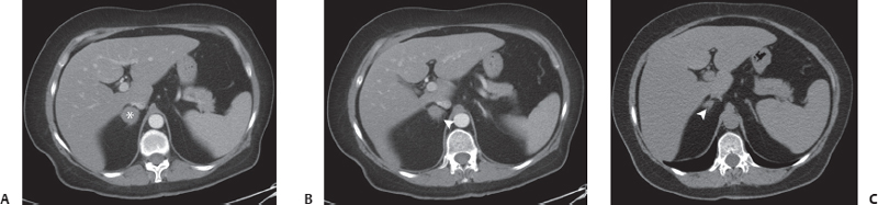
(A) Noncontrast computed tomography (CT) image at the level of the adrenal gland shows a high-attenuation mass (asterisk) in the right adrenal gland. (B) Noncontrast CT at a level lower than that in Figure A shows stranding (arrowhead) in the soft tissues surrounding the right adrenal mass. (C) Noncontrast CT 6 months later at the same level as that in Figure A shows resolution of the right adrenal mass (arrowhead).
 Differential Diagnosis
• Adrenal hemorrhage:
Stay updated, free articles. Join our Telegram channel

Full access? Get Clinical Tree


