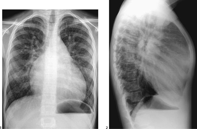
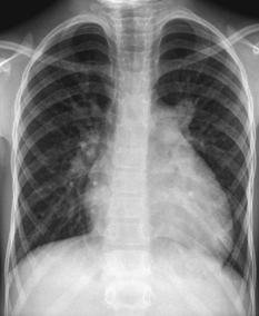
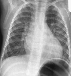
CASE 52 An 11-year-old boy presents with systolic murmur and dyspnea on exertion. Figure 52A A frontal chest radiograph (Fig. 52A1) shows situs solitus and levocardia. The heart is moderately enlarged, and pulmonary vascularity is markedly increased. Increased vascularity is more evident in lateral view (Fig. 52A2) The degree of cardiomegaly reflects the amount of left-to-right shunting. The sternum shows anterior bowing due to cardiomegaly. There is left atrial enlargement. The aortic knob, however, is not prominent. Figure 52B Frontal chest radiograph of a patient with an atrial septal defect shows enlarged heart and increased pulmonary vascularity. There is no evidence of left atrial enlargement. The aortic knob is small. Figure 52C Frontal chest radiograph of a patient with a patent ductus arteriosus shows enlarged heart and increased pulmonary vascularity. The aortic knob is prominent. The enlarged left atrium forms a double contour on the right side. Ventricular septal defect (VSD) Other causes of left-to-right shunt should be considered.
Clinical Presentation

Radiologic Findings


Diagnosis
Differential Diagnosis
Discussion
Clinical Findings
Stay updated, free articles. Join our Telegram channel

Full access? Get Clinical Tree


