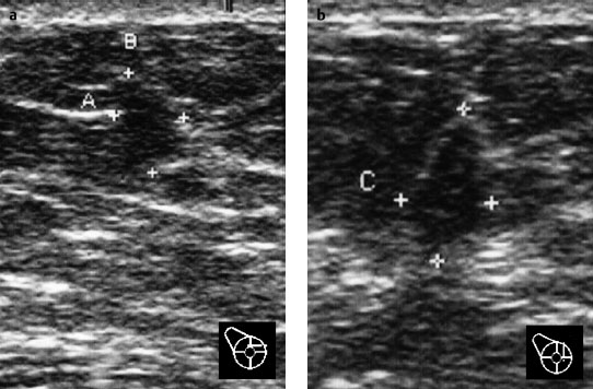
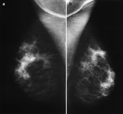
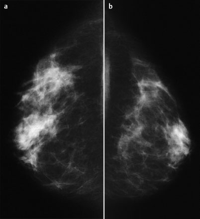
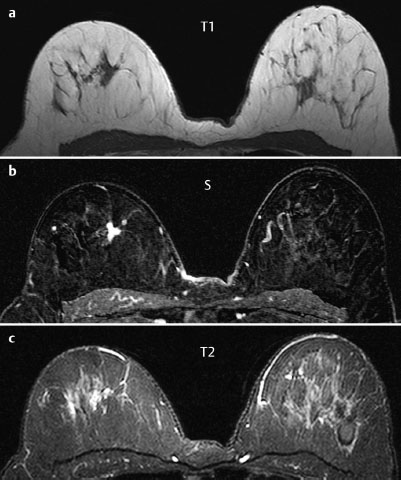
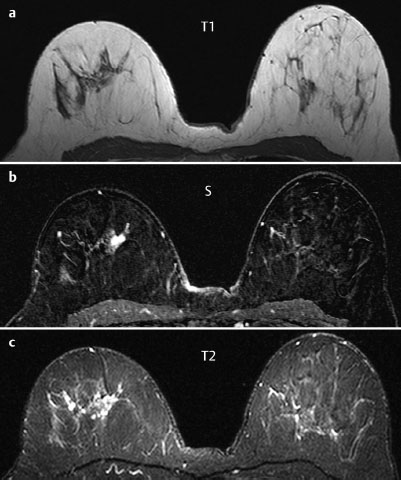
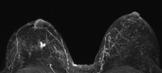
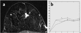
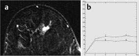
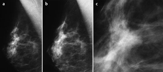
Case 53 Indication: Screening. History: Unremarkable. Risk profile: No increased risk. Age: 52 years. Fig. 53.1 a,b Ultrasound images of outer quadrants of the right breast [imaging not performed by authors]. Nodular parenchymal texture. No circumscribed findings. Fig. 53.2a,b Conventional mammography, MLO view [imaging not performed by authors]. Fig. 53.3a,b Conventional mammography, CC view [imaging not performed by authors]. Fig. 53.4a–c Contrast-enhanced MRI of the breasts. Fig. 53.5a–c Contrast-enhanced MRI of the breasts. Fig. 53.6 Contrast-enhanced MR mammography. Maximum intensity projection. Fig. 53.7a,b Signal-to-time curves. Fig. 53.8a,b Signal-to-time curves. Please characterize ultrasound, mammography, and MRI findings. What is your preliminary diagnosis? What are your next steps? The images presented were taken during a screening examination of an asymptomatic woman. Ultrasound demonstrated a hypoechoic lesion 8 mm in diameter with indeterminate distal echo pattern between the upper quadrants of the right breast. There were also signs of slight architectural distortion. Otherwise, acoustic texture was normal. US BI-RADS right 4/left 1. Mammograms showed partially inhomogeneous dense parenchyma, ACR type 3. In the upper part of the right breast (MLO view) there was a hyperdensity and the resulting asymmetry was visible when both breasts were compared. There were no suspicious masses and no microcalcifications. BI-RADS right 3/left 1. PGMI: CC view P; MLO view G (bilateral axillary skin folds). Additional imaging in a third view (LM) and a magnification mammography of the right breast were also performed (Fig. 53.9a, b,c). Between the upper quadrants of the right breast, a single partially ill-defined, partially spiculated mass with homogeneous enhancement was demonstrated. It showed moderate initial signal increase and a postinitial plateau, and had a slightly increased signal in T2-weighted imaging. The lesion diameter was 15 mm. MRI Artifact Category: 2 MRI Density Type: 1 Fig. 53.9a–c Analogue mammography (LM view), magnification view and enlarged image of the right breast [imaging not performed by authors].
Clinical Findings

Ultrasound
Mammography
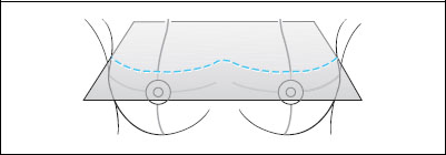
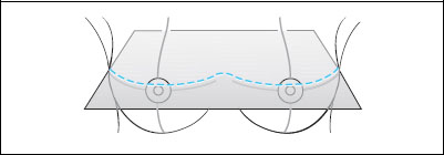
MR Mammography (imaging performed elsewhere)
MRM score | Finding | Points |
Shape | irregular | 1 |
Border | spiculated | 1 |
CM Distribution | inhomogenous | 0 |
Initial Signal Intensity Increase | moderate | 1 |
Post-initial Signal Intensity Character | plateau | 1 |
MRI score (points) |
| 4 |
MRI BI-RADS |
| 4 |
 Differential Diagnosis
Radial scar, invasive carcinoma.
Clinical Findings | right 1 | left 1 |
Ultrasound | right 4 | left 1 |
Mammography | right 4 | left 1 |
MR Mammography | right 4 | left 1 |
BI-RADS Total | right 4 | left 1 |
Procedure
Investigation of the lesion in the right breast by MR-guided vacuum biopsy.
Histopathology of the core biopsy specimen
Tubular carcinoma.
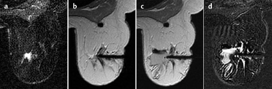
Fig. 53.10a–d MR-guided core biopsy. The lesion in the right breast was reproducible. The coaxial needle is shown in position before the intervention. Precontrast and postcontrast images after removal of specimen are shown.
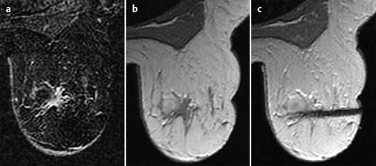
Fig. 53.11 a-c Preoperative MR-guided hook-wire localization before open biopsy.
Histology
Tubular carcinoma.
TC pTlc pN0, G1.
Stay updated, free articles. Join our Telegram channel

Full access? Get Clinical Tree


