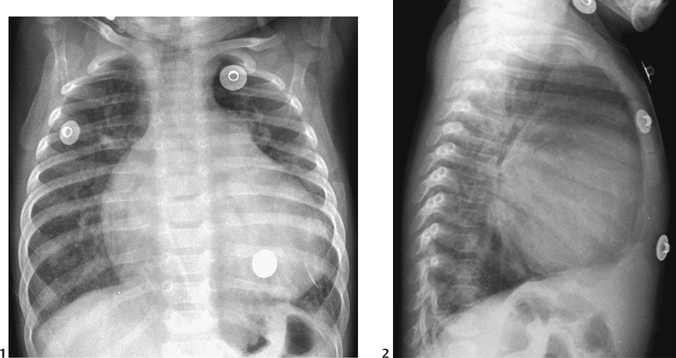
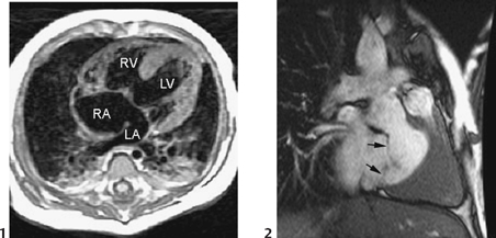
CASE 53 A 5-month-old infant presents with Down syndrome. Figure 53A A frontal chest radiograph (Fig. 53A1) shows situs solitus and levocardia. The heart is moderately enlarged with a globular appearance. The pulmonary vascularity is moderately increased, and pulmonary arterial segment is convex. Both lungs are hyperinflated, with flattening of diaphragms in lateral view (Fig. 53A2). Figure 53B (1) T1-weighted axial MRI shows a large atrioventricular septal defect with a free-floating common atrioventricular valve. LA, left atrium; LV, left ventricle; RA, right atrium; RV, right ventricle. A frame of cine MRI through the ventricular septum (2) called en face image of the ventricular septum, shows a large atrioventricular septal defect and free-floating atrioventricular valve leaflets (arrows). Atrioventricular septal defect. A black-blood MRI in axial plane (Fig. 53B1) shows a large confluent defect involving the atrioventricular junction. A free-floating atrioventricular valve divides the defect into interatrial and interventricular components. A bright-blood MRI through the ventricular septum (Fig. 53B2) shows the scooped-out defect and free-floating atrioventricular valve leaflets (arrows). Babies with Down syndrome should be examined with a high suspicion for congenital heart disease. The overall incidence of congenital heart disease in Down syndrome is —40%. The common cardiac defects include: The clinical manifestation depends on the size of the defect, the presence of an interventricular component of the defect, the severity of atrioventricular valve regurgitation, and the severity of pulmonary vascular disease. Ostium primum defect with minor atrioventricular valve regurgitation is associated with mild symptoms and signs that are similar to those of a simple atrial septal defect. Complete form with severe atrioventricular valve regurgitation develops symptoms and signs of congestive heart failure in early life. Complete form is commonly associated with Down syndrome or, less commonly, other forms of aneuploidy. Atrioventricular septal defect is characterized by a large confluent septal defect in the atrioventricular junction (Fig. 53B
Clinical Presentation

Radiologic Findings

Diagnosis
Differential Diagnosis
Discussion
Clinical Findings
Pathology
![]()
Stay updated, free articles. Join our Telegram channel

Full access? Get Clinical Tree


