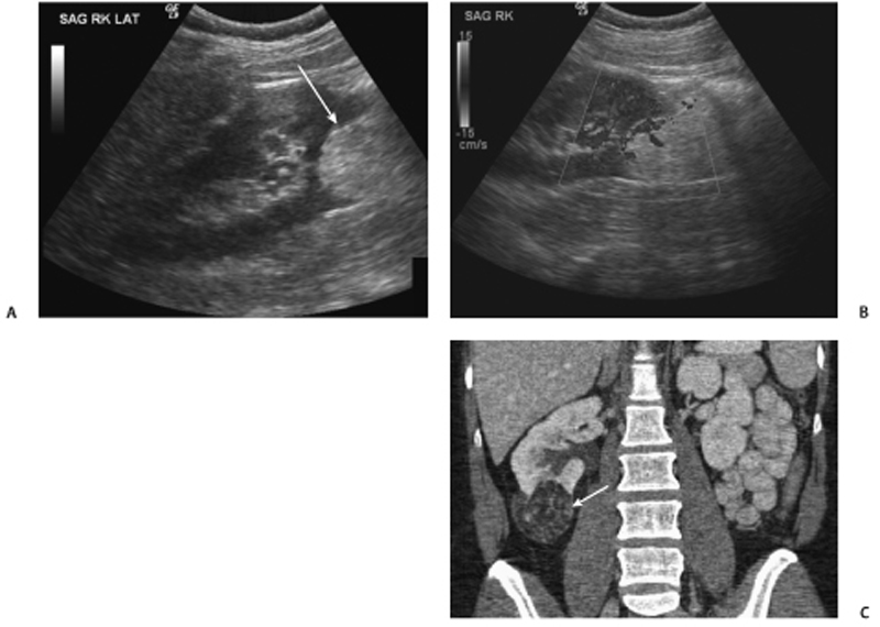
CASE 53 Patient underwent ultrasound exam for nonspecific abdominal pain; incidental finding. Fig. 53.1 (A,B) Ultrasound images show a single large echogenic lesion (arrow) arising from the lower pole of the right kidney with internal color Doppler flow. (C) Contrast-enhanced coronal CT image from the same patient demonstrates a predominantly fat-containing vascular mass in the lower pole of the right kidney (arrow). A sagittal ultrasound image demonstrates a large echogenic lesion in the lower pole of the right kidney (Fig. 53.1A) with internal color Doppler flow (Fig. 53.1B). Contrast-enhanced coronal computed tomography (CT) images from the same patient demonstrate a predominantly fat-containing vascular mass in the lower pole of the right kidney corresponding to the lesion seen on ultrasound (Fig. 53.1C). Renal angiomyolipoma (AML) On ultrasound: On CT:
Clinical Presentation
Radiologic Findings
Diagnosis
Differential Diagnosis
Related posts:


Stay updated, free articles. Join our Telegram channel

Full access? Get Clinical Tree


