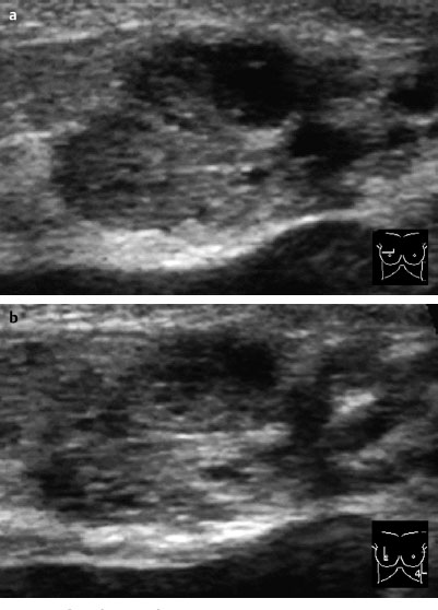
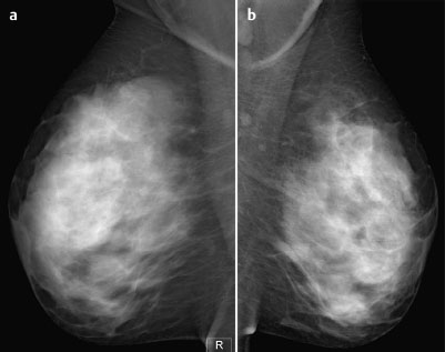
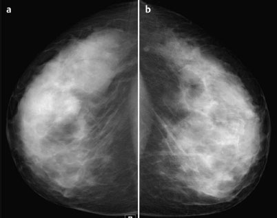
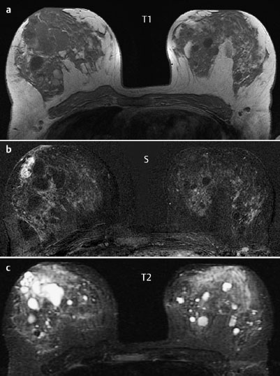
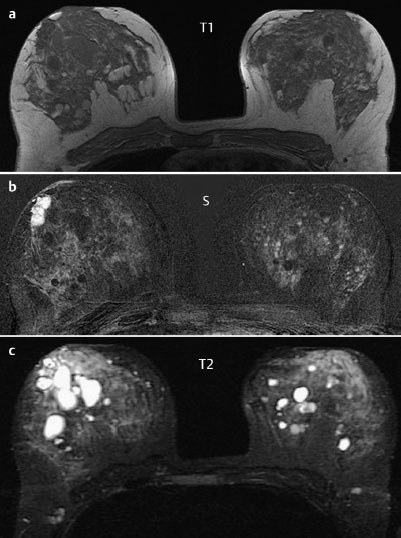
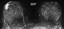
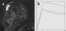
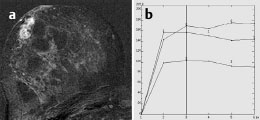
Case 54 Indication: Screening. History: Unremarkable. Risk profile: No increased risk. Age: 44 years. Fig. 54.1 a,b Ultrasound. Dense, nodular parenchyma, but no circumscribed lumps. Fig. 54.2a,b Digital mammography, MLO view. Fig. 54.3a,b Digital mammography, CC view. Fig. 54.4a–c Contrast-enhanced MR mammography. Fig. 54.5a–c Contrast-enhanced MR mammography. Fig. 54.6 Contrast-enhanced MR mammography. Maximum intensity projection. Fig. 54.7a,b Signal-to-time curves. Fig. 54.8a,b Signal-to-time curves. Please characterize ultrasound, mammography, and MRI findings. What is your preliminary diagnosis? What are your next steps? This case illustrates the imaging of an asymptomatic woman in a screening situation. Ultrasound showed an inhomogeneous, partially hypoechoic lesion measuring 3 cm × 1.5 cm in the upper outer quadrant of the right breast. The distal echo pattern was indeterminate. Multiple cysts were also seen in both breasts. US BI-RADS right 3. The parenchyma was bilaterally symmetric and extremely dense, ACR type 4. Under these limiting conditions, mammography showed no suspect densities or masses. No architectural distortion or calcifications. BI-RADS right 1/left 1. PGMI: CC view P; MLO view G (bilateral skin folds). MRI depicted an inhomogeneous enhancing lesion 3 cm × 1.5 cm in size in the upper outer quadrant of the right breast, near the areola. This lesion had very high initial signal increase and postinitial washout. The signal in T2-weighted imaging was partially increased. There were multiple cysts of diameter up to 4 cm in both breasts. MRI Artifact Category: 1 MRI Density Type: 2
Clinical Findings

Ultrasound
Mammography
MR Mammography
MRM score | Finding | Points |
Shape | irregular | 1 |
Border | well-defined | 0 |
CM Distribution | inhomogenous | 1 |
Initial Signal Intensity Increase | strong | 2 |
Post-initial Signal Intensity Character | wash-out | 2 |
MRI score (points) |
| 6 |
MRI BI-RADS |
| 5 |
 Differential Diagnosis
Carcinoma, papilloma, fibroadenoma.
Clinical Findings | right 1 | left 1 |
Ultrasound | right 3 | left 1 |
Mammography | right 1 | left 1 |
MR Mammography | right 5 | left 1 |
BI-RADS Total | right 5 | left 1 |
Procedure
Ultrasound-guided core biopsy of the right breast (Fig. 54.9).
Histology
Intraductal papilloma.
Further Procedure
Open biopsy of the papilloma.
Fig. 54.9a,b US-guided core biopsy (pre-fire, post-fire)
Fig. 54.10a–f MR-guided localization to determine the precise extent of the tumor on the chest wall side. Consecutive subtraction images from the middle of the lesion to its caudal limit (a-d). Precontrast imaging of the tumor at its caudal border (e) and documentation of the correctly positioned hook-wire (f).
Histology
Multifocal minimally invasive papillary carcinoma.
IP pT1mic, pN0(0/10), G2.
Stay updated, free articles. Join our Telegram channel

Full access? Get Clinical Tree


