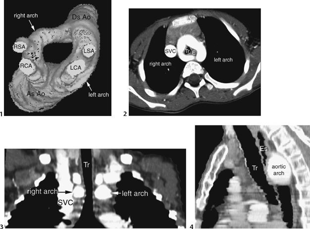
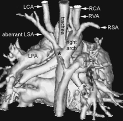
CASE 54 An infant presents with stridor. Figure 54A A frontal chest radiograph (Fig. 54A) shows situs solitus and levocardia. The heart is normal in size and configuration. The pulmonary vascularity is normal. The trachea is not bent to either side and shows concentric narrowing above the carina. The descending parts of aortic knobs are seen in both sides of the distal trachea and spinal column. Figure 54B Three-dimensional CT angiogram (1) shows double aortic arch. AsAo, ascending aorta; DsAo, descending aorta; LCA, left common carotid artery; LSA, left subclavian artery; RCA, right common carotid artery; RSA, right subclavian artery. An axial CT angiogram (2) shows double aortic arch encircling the trachea (Tr) and esophagus (E). SVC, superior vena cava. CT angiogram reconstructed in coronal plane (3) shows concentric narrowing of the trachea (Tr) by compression from the aortic arches. SVC, superior vena cava. CT angiogram reconstructed in sagittal plane (4) shows the large aortic arch compressing the esophagus (Es) from behind. Tr, trachea. (Courtesy of Dr. Yang Min Kim, Seoul, Korea) Double aortic arch. CT angiograms from a different patient (Fig. 54B) show that the vascular ring surrounds the trachea and esophagus. The tracheal compression is well shown in the coronal image, and the esophageal compression in the sagittal image.
Clinical Presentation
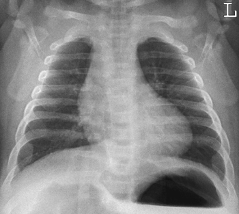
Radiologic Findings
Diagnosis
Differential Diagnosis
![]()
Stay updated, free articles. Join our Telegram channel


Full access? Get Clinical Tree


