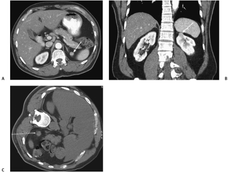
CASE 54 A 72-year-old man presents with a 1-month history of left lower quadrant pain. Fig. 54.1 (A,B) Axial and coronal images from a contrast-enhanced CT demonstrate an exophytic, well-circumscribed enhancing mass (arrows) in the upper pole of the left kidney, which was (C) biopsied under CT guidance. Axial and coronal images from a contrast-enhanced computed tomography (CT) scan demonstrate an exophytic, well-circumscribed enhancing mass in the upper pole of the left kidney (Fig. 54.1A,B), which was biopsied under CT guidance (Fig. 54.1C). Lipid-poor angiomyolipoma
Clinical Presentation
Radiologic Findings
Diagnosis
Differential Diagnosis
Discussion
Background
Related posts:


Stay updated, free articles. Join our Telegram channel

Full access? Get Clinical Tree


