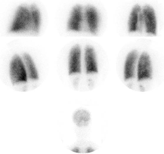
CASE 54 A 67-year-old woman presents with worsening shortness of breath. Fig. 54.1 • A 3.0 mCi dose of 99mTc-MAA is administered intravenously with the patient supine. • The patient should cough and take several deep breaths before administration of the MAA to clear any areas of resting atelectasis. • The patient should breathe normally during tracer injection. • Use a low-energy, all-purpose collimator. • Energy window 20% centered at 140 keV. • Imaging time is 500,000 counts per view. • Matrix size is 128 × 128. • Views are anterior, right anterior oblique, left anterior oblique, posterior, right posterior oblique, and left posterior oblique. Lateral views can also be obtained, although it is important to remember that counts from the contralateral lung will contribute to these views. The lung perfusion images demonstrate overall mildly heterogeneous tracer localization, without segmental defects (Fig. 54.1
Clinical Presentation
Technique
Image Interpretation
![]()
Stay updated, free articles. Join our Telegram channel


Full access? Get Clinical Tree


