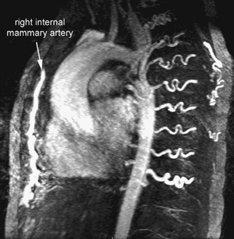
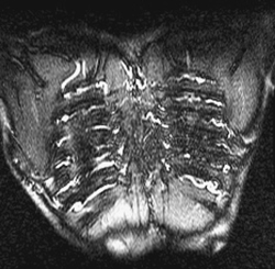
CASE 55 A 12-year-old girl presents with hypertension. Figure 55A A frontal chest radiograph (Fig. 55A) shows situs solitus and levocardia. The heart is mildly enlarged with elongation of the left ventricular border. The prominent ascending aorta forms the right upper heart border. The aortic knob is large for the patient’s age. The proximal descending aorta shows mild indentation, producing a so-called 3 sign. A few ribs show small sclerotic changes along the inferior margins. Figure 55B1 Contrast-enhanced MR angiogram shows discrete coarctation of aorta. Note the collaterals. Figure 55B2 Bright-blood MRI of the posterior wall of the chest of the same patient shows dilated intercostal arteries coursing along the inferior margins of the ribs. Coarctation of the aorta. A contrast-enhanced MR angiogram (Fig. 55B1) in left anterior oblique view from the same patient shows tight discrete stenosis of the aorta at the junction between the isthmic segment of the aortic arch and descending aorta. Notice the dilated intercostal and internal mammary arteries functioning as collateral channels. A bright-blood MRI of the posterior chest wall in coronal plane (Fig. 55B2) shows that the tortuous dilated intercostal arteries course along the inferior borders of the ribs.
Clinical Presentation
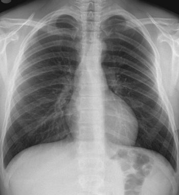
Radiologic Findings
Diagnosis
Differential Diagnosis
Discussion
Clinical Findings
Related posts:


Stay updated, free articles. Join our Telegram channel

Full access? Get Clinical Tree


