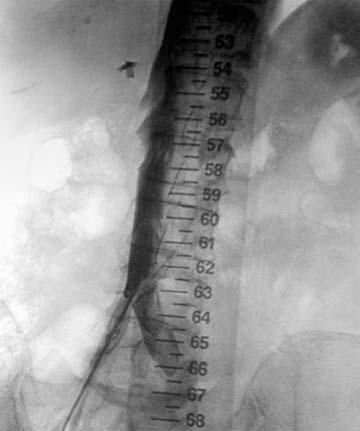
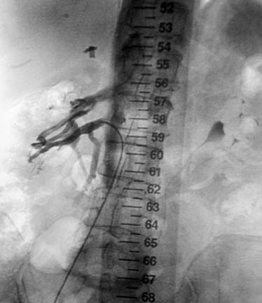
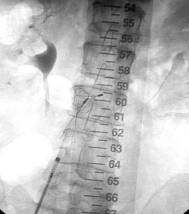
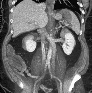
CASE 55 A 62-year-old male status post right total knee arthroplasty complained of left leg swelling with pain behind the knee. Physical exam showed a positive Homans’s sign and duplex sonography revealed deep venous thrombosis (DVT) of the left femoral vein. He was referred to interventional radiology for inferior vena cava (IVC) filter placement. Figure 55-1 A 62-year-old male with right femoral DVT presented to interventional radiology for IVC filter placement. IVC venogram shows inflow from two large right-sided veins. No intraluminal thrombus was visible. The right common femoral vein was punctured with a Micropuncture set (Cook, Bloomington, Indiana) and a 5-French (F) pigtail catheter was placed in the right common iliac vein. An IVC venogram showed inflow from two large right-sided veins. No intraluminal thrombus was visible (Fig. 55-1). Duplicated right renal vein. The pigtail catheter was exchanged over a standard guidewire for a visceral selective catheter, and the lowest right-sided vein was selectively catheterized. Venography revealed two large, communicating renal veins (Fig. 55-2). A Simon Nitinol (Bard Peripheral Vascular, Murray Hill, New Jersey) IVC filter was deployed below the lowest right renal vein (Fig. 55-3). Figure 55-3 A Simon Nitinol filter (Bard Peripheral Vascular, Murray Hill, New Jersey) was deployed below the lowest renal vein. 5F Micropuncture set (Cook, Bloomington, Indiana) 0.035” standard guidewire (15J; Boston Scientific, Natick, Massachusetts) 5F pigtail flush catheter (Cook, Bloomington, Indiana) 5F Visceral Selective catheter (RC-1; Cook, Bloomington, Indiana) Vena cava filter set (Simon Nitinol; Bard Peripheral Vascular, Murray Hill, New Jersey) Anomalies of the renal veins and IVC have been observed by selective renal venography in 20 to 37% of the general population. The most common anomalies include circumaortic left renal vein, retroaortic left renal vein, multiple left or right renal veins, duplication of the IVC, azygous continuation of the IVC, and congenital megacava. The reported incidence rates of these anomalies are as variable as the methods used to detect them, including conventional venography, CT, MRI, cadaveric studies, and observations made during surgical procedures.
Clinical Presentation
Radiologic Studies
Diagnosis
Treatment
Equipment
Discussion
Background
Related posts:


Stay updated, free articles. Join our Telegram channel

Full access? Get Clinical Tree


