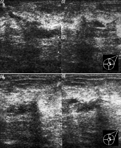
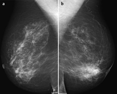
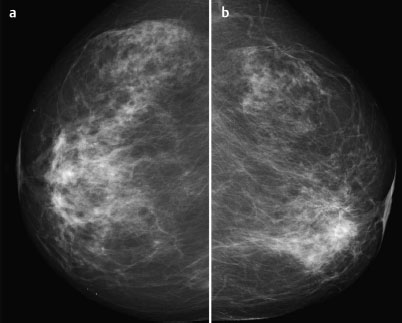
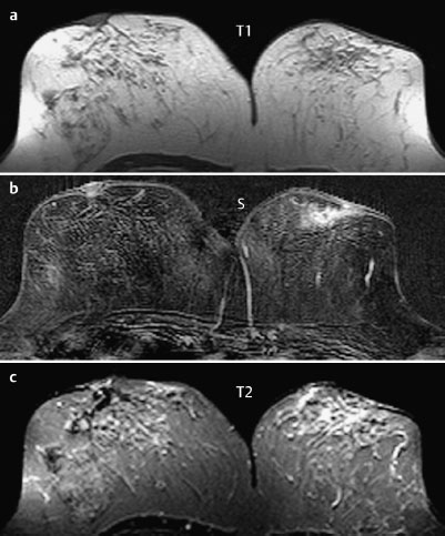
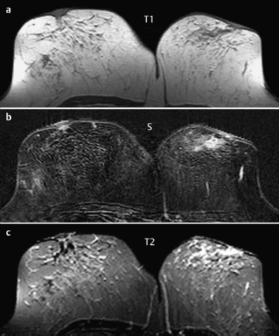
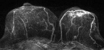
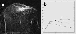
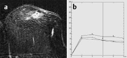
Case 56 Indication: Inflammation of the left breast. History: Unremarkable. Risk profile: No increased risk. Age: 73 years. Fig. 56.1 a,b Ultrasound [imaging not performed by authors]. Painful redness of both upper quadrants of the left breast. No resistance. Fig. 56.2a,b Digital mammography, MLO view [imaging not performed by authors]. Fig. 56.3a,b Digital mammography, CC view [imaging not performed by authors]. Fig. 56.4a–c Contrast-enhanced MRI of the breasts. Fig. 56.5a–c Contrast-enhanced MRI of the breasts. Fig. 56.6 Contrast-enhanced MR mammography. Maximum intensity projection. Fig. 56.7a,b Signal-to-time curves. Fig. 56.8a,b Signal-to-time curves. Please characterize ultrasound, mammography, and MRI findings. What is your preliminary diagnosis? What are your next steps? Imaging was carried out in this case to investigate inflammation of the left breast. A linear hypoechoic region with distal shadowing was depicted in the upper quadrants of the left breast near the nipple. No circumscribed masses. US BI-RADS left 3. The parenchyma had a fibroglandular texture, ACR type 2, and showed asymmetry in mammography with greater density of the left breast than the right. Mammograms showed no masses, no densities, and no microcalcifications. There was a possible thickening of the are olar region in the left breast when compared to the right (caution: digital images). BI-RADS right 1/left 3. PGMI: CC view P; MLO view M (right nipple not in profile, pectoral muscle). An asymmetric enhancement was visible in the region above the left nipple, corresponding to the asymmetry depicted in mammography. It showed ring enhancement, an initial signal increase of 90%, and postinitial washout. Signal in T2-weighted imaging was increased. MRI Artifact Category: 2 MRI Density Type: 1
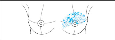
Clinical Findings
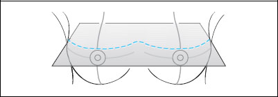

Ultrasound
Mammography
MR Mammography
MRM score | Finding | Points |
Shape | irregular | 1 |
Border | ill-defined | 1 |
CM Distribution | ring | 2 |
Initial Signal Intensity Increase | moderate | 1 |
Post-initial Signal Intensity Character | wash-out | 2 |
MRI score (points) |
| 7 |
MRI BI-RADS |
| 5 |
 Differential Diagnosis
Inflammatory carcinoma, benign inflammation (e.g., nonpuerperal mastitis).
Clinical Findings | right 1 | left 4 |
Ultrasound | right 1 | left 3 |
Mammography | right 1 | left 3 |
MR Mammography | right 1 | left 5 |
BI-RADS Total | right 1 | left 5 |
Procedure
Antibiotic therapy (penicillin) for 10 days. Since this did not result in any improvement in the inflammation, open biopsy was performed to exclude the possibility of inflammatory carcinoma. MR mammography was performed for the purpose of preoperative staging of a potential carcinoma.
Histopathology of the wedge biopsy
Gout (Fig. 56.9).
Fig. 56.9a,b Histological step sections of the manifestation of gout in the breast.
